
This Quick Tips post is the fifth in the series on age estimation on skeletal remains, if you haven’t read the previous post click here, or to start at the beginning click here. The previous post provides an overview of the cranial suture method of aging, whereas the first post covers the basics.
This method is one of the most common ways of chronically aging a human skeleton, and involves examining the surface of the pubis of the os coxae.
Over a lifetime the surface of the pubis change; in early adulthood the surface is rugged and is traversed by horizontal ridges and intervening grooves. By the age of thirty-five, the surface becomes smoother bound by a rim, as it loses relief. The pubic symphysis of an adult over the age of thirty-five, continues to erode and deteriorate with progressive changes.
These changes were first documented by Todd (1920) who conducted a study on 306 males of known age-at-death. Todd identified that there were four parts to the pubic symphysis, where he noted evidence of billowing, ridging, ossific nodules, and texture:
- The ventral border (rampart).
- The dorsal border (rampart).
- The superior extremity.
- The inferior extremity.
Using his observations, Todd identified ten phases of pubic symphysis age, ranging from eight/nine-teen years old to fifty-plus years.

After Todd’s (1920) method which only looked at males, Suchey-Brooks (1990) undertook a study that involved both female and male pubic symphyses – which allowed for a new symphysis scoring system to be created. This new scoring system is made up of six phases, which have a corresponding statistical analysis for the age that each stage represents. The six stages are as follows:
- Lack of delimitation of either superior/inferior extremity; Symphyseal face has a billowing surface (ridges and furrows), which usually extends to include the pubic tubercle. The horizontal ridges are well-marked, and ventral bevelling may be commencing. Although ossific nodules may occur on the either extremity.
- Surface has commencing delimitation of lower and/or upper extremities occurring with or without ossific nodules; Symphyseal face may still show ridge development. The ventral rampart may be in beginning phases as an extension of the bony activity at either or both extremities.
- Ventral rampart in process of completion; There can be a continuation of fusing ossific nodules forming the upper extremity and along the vetral border. Symphyseal face is smooth or can continue to show distinct ridges. Dorsal plateau is complete. Absence of lipping of symphyseal dorsal margin; no bony ligamentous outgrowths.
- Oval outline is complete, but a hiatus can occur in upper ventral rim; Symphyseal face is generally fine grained although remnants of the old ridge and furrow system may still remain. Pubic tubercle is fully separated from the symphyseal face by definition of the upper extremity. The symphyseal face may have a distinct rim. Ventrally, bony ligamentous outgrowths may occur on inferior portion of pubic bone adjacent to symphyseal face. If any lipping occurs, it will be slight and located on the dorsal border.
- Symphyseal face is completely rimmed with some slight depression of the face itself, relative to the rim; Moderate lipping is usually found on the dorsal border with more prominent ligamentous outgrowths on the ventral border. There is little or no rim erosion. Breakdown may occur on superior ventral border.
- Symphyseal face may show on-going depression as rim erodes; Ventral ligamentous attachments are marked. In many individuals the pubic tubercle appears as a separate bony knob. The face may be pitted or porous, giving an appearance of disfigurement with the on-going process of erratic ossification. Crenulations may occur. The shape of the face is often irregular at this stage.

Figure 2: The Suchey-Brooks pubic symphasis scoring system of the six stages. It is recommended that these illustrations be supplemented by casts before actual aging is attempted.
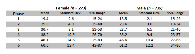
Table 1: Statistics for the Suchey-Brooks phases in females and males.
This pubis symphyseal surface method is often preferred over the other aging methods due to the age-related changes on the pubis surface continuing after full adult stature has occurred, for example; epiphyseal closing method can only age early adulthood.
References:
Buikstra, J.E., Ubelaker, D.H. 1994. Standards for Data Collection From Human Skeletal Remains.Fayetteville, Arkansas: Arkansas Archaeological Survey Report Number 44.
Todd, T.W. 1920 Age changes in the pubic bone: I. The white male pubis. American Journal of Physical Anthropology, 3: 467-470.
White, T.D., Folkens, P.A. 2005. The Human Bone Manual. San Diego, CA: Academic Press. Pg 360-385.
If you’re new to the realm of archaeological, anthropological and forensic sciences (AAFS), or are a student needing sturdy and reliable references, or wondering “what archaeology or anthropology textbooks to buy?” Check out our new ‘Useful Literature’ page!


Pingback: Quick Tips: How To Estimate The Chronological Age Of A Human Skeleton – The Basics. | All Things AAFS!
Reblogged this on devils and black sheep..
Pingback: Quick Tips: How To Estimate The Chronological Age Of A Human Skeleton – Pubic Symphyseal Surface | Research Group FB Ancient History
Reblogged this on Research Group FB Ancient History.
Pingback: Quick Tips: How To Estimate The Chronological Age Of A Human Skeleton – Sternal Rib End Method. – All Things AAFS!