
This is the 4th blog post in this Quick Tips series on chronologically dating human skeletal remains, if you haven’t read the first post click here to start at the beginning. In my previous blog post I introduced the method of chronologically dating sub-adults using dentition, you can find out this information by clicking here.
Another method of chronologically aging human skeletal remains is by observing the cranial suture closure sites. The human skull has seventeen unique cranial fusion sites (Figure 1), that are positioned on the vault, the lateral-anterior sites, and the maxillary suture. The seventeen sites are:
- Midlambdoid 10.Superior sphenotemporal
- Lambda 11. Incisive suture
- Obelion 12. Anterior median palatine
- Anterior sagittal 13. Posterior median palatine
- Bregma 14. Transverse palatine
- Midcoronal 15. Sagittal (endocranial)
- Pterion 16. Left lambdoidal (endocranial)
- Sphenofrontal 17.Left coronal (endocranial)
- Inferior sphenotemporal
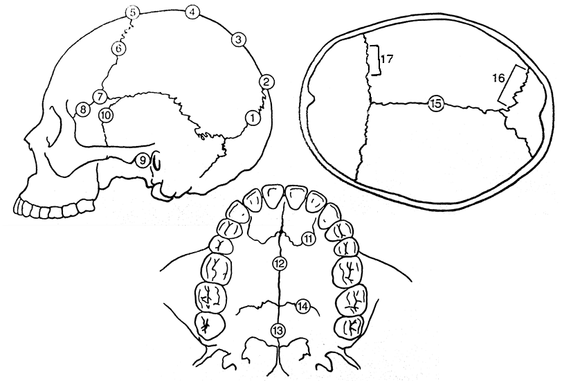
Figure 1) Diagram showing the seventeen cranial suture sites.
The first seven fusion sites are on the vault, and the lateral-anterior sites consist of numbers six to ten. Each suture is usually given a numerical score, the score of 0-3 is recommended by the Buikstra and Ubelaker standards (1994). The Buikstra and Ubelaker (1994) scoring system is as follows;
- 0 is given when the suture is open, meaning there is no evidence of ectocranial closure.
- 1 is given where there is a minimal closure of the suture.
- 2 is given to sutures with evidence of significant closure.
- 3 is given to a completely obliterated suture (complete fusion).
So to attain the age of a skeletal remain you would total the scores for each grouping of sites, vault (1-7) or lateral anterior (6-10), and by comparing the scores to the known composite scores vs. chronological age of Meindl And Lovejoy, 1985 (Figure 2).

Figure 2: Table demonstrating Meindl and Lovejoy (1985)’s composite scores of the sutures on the vault and lateral-anterior, respectively, in relation to mean chronological age.
A very useful cranial suture site is the sphenooccipital synchrondrosis, because at least 95% of all individuals have fusion here between the ages of twenty and twenty-five, with most individuals experiencing complete fusion around the age of twenty-three (Krogman and Işcan, 1986).
References:
Buikstra, J.E., Ubelaker, D.H. 1994. Standards for Data Collection From Human Skeletal Remains.Fayetteville, Arkansas: Arkansas Archaeological Survey Report Number 44.
Krogman, W.M., Işcan, M.Y. 1986. The Human Skeleton in Forensic Medicine (2nd Ed). Springfield, Illinois: C.C. Thomas.
Meindl, R.S., Lovejoy, C.O. 1985. Ectocranial Suture Closure: A Revised Method For The Determination Of Skeletal Age At Death Based On The Lateral-Anterior Sutures. American Journal of Physical Anthropology. 68, 57-66.
White, T.D., Folkens, P.A. 2005. The Human Bone Manual. San Diego, CA: Academic Press. Pg 360-385.
This is the forth of a Quick Tips series on ageing skeletal remains, the next in this series will focus on the use of the pubic symphyseal surface to chronologically age skeletal remains. To read more Quick Tips in the meantime, click here!
To learn about basic fracture types and their characteristics/origins in their own Quick Tips series, click here!


Reblogged this on Virtual Curation Museum and commented:
Very informative and great for my classes!
Pingback: Quick Tips: How To Estimate The Chronological Age Of A Human Skeleton – The Basics. | All Things AAFS!
Pingback: Quick Tips: How To Estimate The Chronological Age Of A Human Skeleton – Pubic Symphyseal Surface Method. | All Things AAFS!