Quick Tips – Common Questions: Why are some diseases more easily identified on skeletal remains than others?

This is a Quick Tips post providing a basic answer to a commonly asked question often faced within the field of archaeology and anthropology.

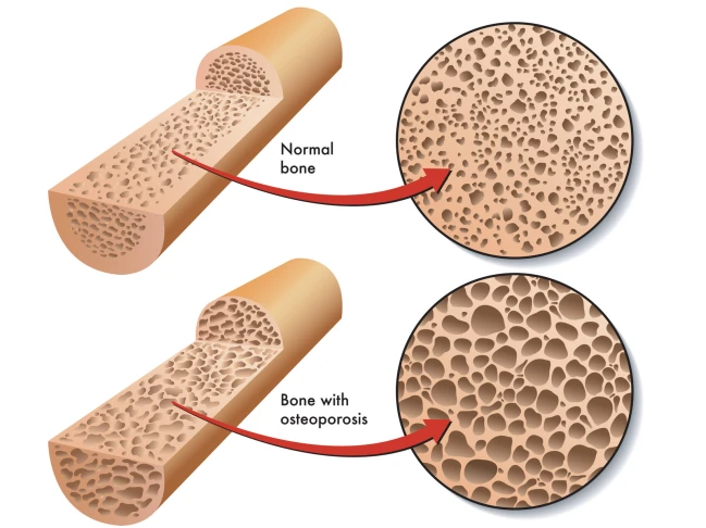
Some diseases are more easily to identify on skeletal remains due to leaving tell-tale signs in the bones preservation. An easy example of this is osteoporosis; this condition leaves the inners of bones a lot more porous which is easier to visually assess and compare to a ‘healthy’ individual’s skeletal remains.
canstockphoto18602582

Some diseases are more easily to identify on skeletal remains due to leaving tell-tale signs in the bones preservation. An  example of this is osteoporosis; this condition leaves the inners of bones a lot more porous than normal bones.

A study by Hershkovitz & Rothschild (1997) highlighted how certain medical conditions, in their study sickle cell anaemia, affects the bone growth and development. Hershkovitz & Rothschild found that due to the iron deficiency from sickle cell anaemia caused porotic hyperostosis (symmetrical osteoporosis) on the parietal bone as well as others. They were able to visually diagnose this due to the characteristic ‘pores’ over the skull.

Another example of an easily identifiable disease is tuberculosis (TB), TB can cause devastating bone damage. A recent archaeological study by Lewis (2011) looked into a population who suffered from TB. Lewis visually analysed the skeletal remains of a juvenile population from Poundbury Camp, Dorset. The TB infection caused numerous ailments to the infected, such as fever, but it’s the skeletal damage which gave the indication that the person suffered.  Amongst the population there was a high instance of skeletons with necrosis and lytic lesions characterised by minimal bone formation.  Many of the juvenile’s vertebrae displayed new bone formations which could indicate the presence of a paravertebral abscess. Many of the metatarsals were also displaying evidence of new bone formation which they concluded could be indicative of tuberculous dactylitis. Osteomyelitis, infection of the bone, was also found on a few mandibles and visually diagnosed due to its characteristic small pores found in a localised area. It is this characterised skeletal damage, seen on numerous cases during known TB outbreaks, which cause more diseases to be easily identified by eye due to the skeletal anomalies.

There are problems when trying to differentiate certain diseases for example; TB with brucellosis (undulant fever). As they both produce spinal lesions it is necessary to observe the other characteristic skeletal damage (new bone formation and osteomyelitis) to correctly identify it as a TB infection. Another slight difference between TB and brucellosis is that the spinal lesions are more sclerotic and regular than those from a TB infection (Lewis, 2011).

These porous bones and unexpected bone formations are easily observed, as they are not what’s expected during the known skeletal development found in healthy persons. Problems arise with diseases that do no damage to the skeleton, but instead affect soft tissue and muscles. These illnesses are harder to identify as they decay over time leaving only trace elements in the surrounding soils which would then hold the key for disease identification.

References:

Hershkovitz, I. Rothschild, B. et al. 1997. Recognition of sickle cell anemia in skeletal remains of children. American Journal of Physical Anthropology. Volume 104, Issue 2. 213-226.

Lewis, M. 2011. Tuberculosis in the non-adults from Romano-British Poundbury Camp, Dorset, England. International Journal of Paleopathology. Volume 1, Issue 1. 12-23.

To learn how archaeologists and anthropologists use teeth to age skeletal remains, read our Quick Tips: How To Estimate The Chronological Age of a Human Skeleton – Using Dentition to Age Subadults. Or to read more of our interesting Quick Tips, click here.

Textbook of the Week: Forensic Archaeology Advances in Theory and Practice.

Every week we highlight one archaeology/anthropology textbook from our suggested readings, a full list of our suggested resources can be found here, on our Useful Literature page.

5191V5QW55L._SY344_BO1,204,203,200_

Forensic Archaeology Advances in Theory and Practice (UK/Europe Link)
Forensic Archaeology Advances in Theory and Practice (US/Worldwide Link)
by John Hunter & Margaret Cox. Rating: ****

“This text book is easy to follow, so perfect for beginners or first year students. It uses numerous case studies and illustrations to show you how to apply it in practice, meaning that you can fully grasp what situation to use it in and how to correctly apply it.

If you’re a student – check out our ‘Quick Tips’ posts where we breakdown topics of AAFS into bite-sized chunks. We’re currently covering how to age and how to estimate the biological sex of skeletal remains, and also how to identify dental diseases!

Quick Tips – Common Questions: What can an anthropologist tell from the examination of teeth regarding either forensic identification of individuals or understanding past populations?

This is a Quick Tips post providing a basic answer to a commonly asked question often faced within the field of archaeology and anthropology.

An anthropologist can obtain a wide and varied collection of information from examining teeth. Information such as paleodiets and palaeoenvironments can be learnt from studying a population, or from studying an individual sample you can identify how old the person was at time of death or whether that person was pregnant/ill. These examples are just the tip of the iceberg on what you can learn from dentition.

Ondontology

An anthropologist can obtain a wide and varied collection of information from examining teeth, ranging from palaeodiets and palaeoenvironmental information to age of death.

From studying a large population dentition sample, a picture can be painted of their past diets, current diets and palaeoenvironments. Isotopes play a huge part in conducting research into palaeodiets and palaeoenvironments.

Isotopes are deposited into the teeth of an individual/population from food sources or environment. A tooth can provide isotopic information from the past 20yrs of the individual’s life. The enamel and dentine can be examined to analyse the isotopic values that will pinpoint an origin of a population or food sources. The carbon and nitrogen isotope compositions found within the enamel are used to reconstruct diet and the oxygen isotopes are used to determine the geographic origin of the food source. The carbon isotopes are absorbed from the diet of the animals that are sources and the oxygen isotopes from the water that the population consume. These isotopic values are vital in helping an anthropologist understand the local ecosystem a population exploited and whether a population migrated to numerous locations which caused changes in the available diet.

The cementum of a tooth can highlight important information about a person which can be used for forensic identification; this information could give an approximate age of death. An example of this application is seen in Kagerer and Grupe (2000) study where they obtained 80 freshly extracted teeth and investigated the incremental lines in acellular extrinsic fibre cementum. From studying the cementum, they were able to determine the age of the patient by comparing it to detailed queries of the patients life history. This study also identified patients who were pregnant. Kagerer and Grupe (2000) concluded that if there was a presence of hypo-mineralised incremental lines on the extracted tooth, the patient was pregnant. This is due to the pregnancies influence on calcium metabolism. A confliction with this is that hypo-mineralized lines can also appear when a skeletal trauma or renal illness was present.

By looking at the dentition of molars the age of the skeleton can be estimated. A recent study by Mesotten, et al. (2002) highlighted the application of forensic odontology. Mesotten, et al’s methodology consisted of examining 1175 orthopantomograms which belonged to patients who were of Caucasian origin and were aged between 16 and 22years. From their investigation Mesotten, et al. were able to conclude that from studying the molars, it was possible to age Caucasian individuals with a regression formula with a standard deviation of 1.52 or 1.56 years for males and females, respectively, if all four third molars were available. This could play a fundamental role in identifying a missing person by estimating the decease’s age and seeing if its estimate matches the individual.

Although the studies from Mesotten, et al (2002) and Kagerer and Grupe (2000) have been written about and applied to individual cases, their methodology and conclusions can be applied to a past population if a group of skeletons were found with preserved teeth. The individual’s age of death can be used as quantitative data, alongside other individuals from the same sample, to figure out a past population’s life expectancy.

References:

Kagerer, P. Grupe, G. 2000. Age-at-death diagnosis and determination of life-history parameters by incremental lines in human dental cementum as an identification aid. Forensic Science International. 118, 1. 75-82.

Mesotten, K. Gunst, K. Carbonez, A. Willems, G. 2002. Dental age estimation and third molars: a preliminary study. Forensic Science International. Volume 129, Issue 2, 110-115

To learn how archaeologists and anthropologists use teeth to age skeletal remains, read our Quick Tips: How To Estimate The Chronological Age of a Human Skeleton – Using Dentition to Age Subadults. Or to read more of our interesting Quick Tips, click here.

Quick Tips: How To Estimate The Chronological Age Of A Human Skeleton – Pubic Symphyseal Surface Method.

This Quick Tips post is the fifth in the series on age estimation on skeletal remains, if you haven’t read the previous post click here, or to start at the beginning click here. The previous post provides an overview of the cranial suture method of aging, whereas the first post covers the basics.

This method is one of the most common ways of chronically aging a human skeleton, and involves examining the surface of the pubis of the os coxae.

Over a lifetime the surface of the pubis change; in early adulthood the surface is rugged and is traversed by horizontal ridges and intervening grooves. By the age of thirty-five, the surface becomes smoother bound by a rim, as it loses relief. The pubic symphysis of an adult over the age of thirty-five, continues to erode and deteriorate with progressive changes.

These changes were first documented by Todd (1920) who conducted a study on 306 males of known age-at-death. Todd identified that there were four parts to the pubic symphysis, where he noted evidence of billowing, ridging, ossific nodules, and texture:

  1. The ventral border (rampart).
  2. The dorsal border (rampart).
  3. The superior extremity.
  4. The inferior extremity.

Using his observations, Todd identified ten phases of pubic symphysis age, ranging from eight/nine-teen years old to fifty-plus years.

Image

After Todd’s (1920) method which only looked at males, Suchey-Brooks (1990) undertook a study that involved both female and male pubic symphyses – which allowed for a new symphysis scoring system to be created. This new scoring system is made up of six phases, which have a corresponding statistical analysis for the age that each stage represents. The six stages are as follows:

  1. Lack of delimitation of either superior/inferior extremity; Symphyseal face has a billowing surface (ridges and furrows), which usually extends to include the pubic tubercle. The horizontal ridges are well-marked, and ventral bevelling may be commencing. Although ossific nodules may occur on the either extremity.
  2. Surface has commencing delimitation of lower and/or upper extremities occurring with or without ossific nodules; Symphyseal face may still show ridge development. The ventral rampart may be in beginning phases as an extension of the bony activity at either or both extremities.
  3. Ventral rampart in process of completion; There can be a continuation of fusing ossific nodules forming the upper extremity and along the vetral border. Symphyseal face is smooth or can continue to show distinct ridges. Dorsal plateau is complete. Absence of lipping of symphyseal dorsal margin; no bony ligamentous outgrowths.
  4. Oval outline is complete, but a hiatus can occur in upper ventral rim; Symphyseal face is generally fine grained although remnants of the old ridge and furrow system may still remain. Pubic tubercle is fully separated from the symphyseal face by definition of the upper extremity. The symphyseal face may have a distinct rim. Ventrally, bony ligamentous outgrowths may occur on inferior portion of pubic bone adjacent to symphyseal face. If any lipping occurs, it will be slight and located on the dorsal border.
  5. Symphyseal face is completely rimmed with some slight depression of the face itself, relative to the rim; Moderate lipping is usually found on the dorsal border with more prominent ligamentous outgrowths on the ventral border. There is little or no rim erosion. Breakdown may occur on superior ventral border.
  6. Symphyseal face may show on-going depression as rim erodes; Ventral ligamentous attachments are marked. In many individuals the pubic tubercle appears as a separate bony knob. The face may be pitted or porous, giving an appearance of disfigurement with the on-going process of erratic ossification. Crenulations may occur. The shape of the face is often irregular at this stage.
Image

Figure 2: The Suchey-Brooks pubic symphasis scoring system of the six stages. It is recommended that these illustrations be supplemented by casts before actual aging is attempted.

Image

Table 1: Statistics for the Suchey-Brooks phases in females and males.

This pubis symphyseal surface method is often preferred over the other aging methods due to the age-related changes on the pubis surface continuing after full adult stature has occurred, for example; epiphyseal closing method can only age early adulthood.

References:

Buikstra, J.E., Ubelaker, D.H. 1994. Standards for Data Collection From Human Skeletal Remains.Fayetteville, Arkansas: Arkansas Archaeological Survey Report Number 44.

Todd, T.W. 1920 Age changes in the pubic bone: I. The white male pubis. American Journal of Physical Anthropology, 3: 467-470.

White, T.D., Folkens, P.A. 2005. The Human Bone Manual. San Diego, CA: Academic Press. Pg 360-385.

If you’re new to the realm of archaeological, anthropological and forensic sciences (AAFS), or are a student needing sturdy and reliable references, or wondering “what archaeology or anthropology textbooks to buy? Check out our new ‘Useful Literature’ page!

Image

Useful Literature.

Image

If you’re new to the realm of archaeological, anthropological and forensic sciences (AAFS), or are a student needing sturdy and reliable references, or wondering “what archaeology or anthropology textbooks to buy?”
On our ‘Useful Literature’ page you can find links to the full selection of the best textbooks – most of these have been suggested to me by my university Professors.

Here is a short-list of the most helpful student books for archaeology and anthropology:

Quick Tips: How To Estimate The Chronological Age Of A Human Skeleton – Cranial Suture Closure Method.

This is the 4th blog post in this Quick Tips series on chronologically dating human skeletal remains, if you haven’t read the first post click here to start at the beginning. In my previous blog post I introduced the method of chronologically dating sub-adults using dentition, you can find out this information by clicking here.

Another method of chronologically aging human skeletal remains is by observing the cranial suture closure sites. The human skull has seventeen unique cranial fusion sites (Figure 1), that are positioned on the vault, the lateral-anterior sites, and the maxillary suture. The seventeen sites are:

  1. Midlambdoid                                           10.Superior sphenotemporal
  2. Lambda                                                    11. Incisive suture
  3. Obelion                                                    12. Anterior median palatine
  4. Anterior sagittal                                      13. Posterior median palatine
  5. Bregma                                                    14. Transverse palatine
  6. Midcoronal                                              15. Sagittal (endocranial)
  7. Pterion                                                     16. Left lambdoidal (endocranial)
  8. Sphenofrontal                                         17.Left coronal (endocranial)
  9. Inferior sphenotemporal
Image

Figure 1) Diagram showing the seventeen cranial suture sites.

The first seven fusion sites are on the vault, and the lateral-anterior sites consist of numbers six to ten. Each suture is usually given a numerical score, the score of 0-3 is recommended by the Buikstra and Ubelaker standards (1994). The Buikstra and Ubelaker (1994) scoring system is as follows;

  • 0 is given when the suture is open, meaning there is no evidence of ectocranial closure.
  • 1 is given where there is a minimal closure of the suture.
  • 2 is given to sutures with evidence of significant closure.
  • 3 is given to a completely obliterated suture (complete fusion).

So to attain the age of a skeletal remain you would total the scores for each grouping of sites, vault (1-7) or lateral anterior (6-10), and by comparing the scores to the known composite scores vs. chronological age of Meindl And Lovejoy, 1985 (Figure 2).

Image

Figure 2: Table demonstrating Meindl and Lovejoy (1985)’s composite scores of the sutures on the vault and lateral-anterior, respectively, in relation to mean chronological age.

A very useful cranial suture site is the sphenooccipital synchrondrosis, because at least 95% of all individuals have fusion here between the ages of twenty and twenty-five, with most individuals experiencing complete fusion around the age of twenty-three (Krogman and Işcan, 1986).

References:

Buikstra, J.E., Ubelaker, D.H. 1994. Standards for Data Collection From Human Skeletal Remains.Fayetteville, Arkansas: Arkansas Archaeological Survey Report Number 44.

Krogman, W.M., Işcan, M.Y. 1986. The Human Skeleton in Forensic Medicine (2nd Ed). Springfield, Illinois: C.C. Thomas.

Meindl, R.S., Lovejoy, C.O. 1985. Ectocranial Suture Closure: A Revised Method For The Determination Of Skeletal Age At Death Based On The Lateral-Anterior Sutures. American Journal of Physical Anthropology. 68, 57-66.

White, T.D., Folkens, P.A. 2005. The Human Bone Manual. San Diego, CA: Academic Press. Pg 360-385.

This is the forth of a Quick Tips series on ageing skeletal remains, the next in this series will focus on the use of the pubic symphyseal surface to chronologically age skeletal remains. To read more Quick Tips in the meantime, click here

To learn about basic fracture types and their characteristics/origins in their own Quick Tips series, click here!

Share Post Sign

Quick Tips: Forensic Entomology – An Introduction.

What is forensic entomology? It is a discipline within forensic sciences where specialists use information that they know about insect lifecycles and behaviours to interpret evidence in a legal context, relating to humans and animals. Entomologists don’t just stick to insects; their work can expand to include other arthropods, mites, spiders and macro-invertebrates.

Image

Insect species which are relevant to forensic entomology

What information can we learn from insect activity? Insects are everywhere and can hardly be avoided, so it’s no surprise that sometimes they get mixed up in the evidence left behind – making them extremely valuable to an investigation. Insects can be a vital part of forensic science as they can provide a time and date to a crime or even a geographical position to where it happened. As some insects only become apparent during certain months, they can become a biological calendar for when a crime might have been committed. As well as being a biological calendar, certain insect species are only native in specific countries or hemispheres. This can be used to create an ‘X marks the spot’ on where a crime was committed – even if a body was moved/buried. Because of this, insects can be the key to past and present events as well as the future.

The insects that are particularly relevant to forensic entomological investigations are blow flies (diptera), flesh flies, cheese skippers, hide and skin beetles, rove beetles and clown beetles. These forensically relevant insects can be placed in four categories:

  • Necrophages, which feed only on the decomposing tissue of the body or body parts. This is the category that blow flies, hide beetles and clown beetles are classed under.
  • Predators of the necrophages – for example the rove beetles and ground beetles.
  • Omnivores that consume both the live insects inhabiting the corpse and the dead flesh – ants and wasps.
  • Opportunist species, which arrive because the corpse is a part of their local environment. This is where mites, hoverflies, butterflies and occasionally spiders are classified.

Forensic entomologists use the evidence they gain from studying insects within legal cases in either civil or criminal courts. Civil court cases include:

  • Insect infestation in urban contexts.
  • Stored product infestations/pests.

Criminal court cases include:

  • Neglect – either animal or human (elderly and children).
  • Insect infestation of a body – living or dead.
  • Death in which foul play is suspected.

This is just an introduction into the world of forensic entomology, if you’d like to know more or further your knowledge on this topic check out this book, I found it very interesting and a terrific read:

  • Forensic Entomology: An Introduction (UK/Europe)
    Forensic Entomology: An Introduction (US/Worldwide Link)
    by Dorothy Gennard. Rating – ***
    “I used my this for my blog post on the basics of forensic entomology. It is perfect if you’re unsure on whether or not you want to pursue this career/discipline. Definitely a good read if your interest is sparked by Dr Hodgins from ‘Bones’, as it explained everything involved within entomology under legal settings.”

Quick Tips: How To Estimate The Chronological Age Of A Human Skeleton – The Basics.

Estimation of age-at-death involves observing morphological features in the skeletal remains, comparing the information with changes recorded for recent populations of known age, and then estimating any sources of variability likely to exist between the prehistoric and the recent population furnishing the documented data. This third step is seldom recognized or discussed in osteological studies, but it represents a significant element. – Ubelaker, D. 1989.

There are numerous markers on a human skeleton which can provide archaeologists and anthropologists with an estimate age of the deceased. The areas of the skeletal remains that are studied are:

If the skeletal marker listed above is a link, it means that I have already covered it in an individual blog post and can be found by following the link.

We can age skeletal remains to a rough estimate, as over a lifetime a human skeleton undergoes sequential chronological changes. Teeth appear and bone epiphyseal form and fuse during childhood and adolescence, with some bone fusing, metamorphose and degeneration carrying on after the age of twenty. Buikstra and Ubelaker, 1994, developed seven age categories that human osteological remains are separated into. The seven age classes are; fetus (before birth), infant (0-3 years), child (3-12 years), adolescent (12-20 years), young adult (20-35 years), middle adult (35-50 years), and old adult (50+ years).

When it comes to ageing skeletal remains, there are numerous problems. This is because individuals of the same chronological age can show difference degrees of development. Therefore, this causes archaeologists and anthropologists to obtain an accurate age estimate, which may not be precise.

It should be noted that it is a lot easier to deduce a juvenile/sub-adult’s age, as the ends of the limb bones form and fuse at known ages and the ages of which tooth formation and eruption occur are very well documented, although somewhat variable. After maturity there is little continuing skeletal change to observe, this causes adult ageing to become more difficult.

References:

Buikstra, J.E., Ubelaker, D.H. 1994. Standards for Data Collection From Human Skeletal Remains. Fayetteville, Arkansas: Arkansas Archaeological Survey Report Number 44.

Ubelaker, D.H. 1989. Human Skeletal Remains: Excavation, Analysis, Interpretation (2nd Ed.). Washington, DC: Taraxacum.

White, T.D., Folkens, P.A. 2005. The Human Bone Manual. San Diego, CA: Academic Press. Pg 360-385.

This is the first of a Quick Tips series on ageing skeletal remains, the next in this series will focus on the epiphyseal closure method of ageing sub-adults. To read more Quick Tips in the mean time, click here

To learn about basic fracture types and their characteristics/origins click here!

Quick Tips: The Use of 3D Animation to Visualise a Crime Scene in Forensics.

Many television programs create 3D animations and computer generated images using highly technical computer programmes to help re-enact the scenes or time frame of a crime. This is mostly used so that the viewer at home can really grasp what crime has been committed and help establish a sense that they are a witness. But in reality these animations and images are becoming an increasingly popular technique used within the courtroom.

Information and evidence can be easily constructed from the traditional methods of forensic photography, blood spatter analysis and eye witness testimonies. But in this modern technological time the information gathered is now being used to create computerised animation that depicts the series of events within a crime. But is this method of providing visual appropriate and correct? Could the animation be showing a display of actions/movements that humans can’t possibly and physically make?

There is a big issue with admissibility, which can cause bias. This occurs when the jurors or judge aren’t aware of an error/uncertainty within the procedure of recreating a real life scene into animation. This can cause them to believe that the evidence is a hundred per cent correct, when in fact there are many errors which were created in the process or animation (Ma & Zheng, 2010). Another big problem arises when studies found that people are five times more likely to remember something they see and hear rather than hearing alone. People are also twice as likely to be persuaded if the arguments are backed with visual evidence (Lederer & Solomon, 1997). So this poses a huge problem as false memories and false testimonies could be influenced, which in the end could cause an innocent person to go to jail for a crime they did not commit.

So with the possibility of creating false memories is the use of 3D animation beneficial for the use of visualising crime scenes within court? It is argued that it is as the use of computerised images creates a higher level of accuracy and speeds up the forensic investigational process but only in major crime types, not every day homicides and robberies. However even though it has limited application in the courtrooms,  it can pose to be very useful in formal briefs with the forensic personnel, and within the backstage elements of the investigation itself (Ma & Zheng, 2010).

References:

Lederer FI, Solomon SH. 1997. Courtroom technology – an introduction to the onrushing future. Fifth National Court Technology Conference: National Centre for State Courts. Available here.

Ma M, Zheng H. 2010. Virtual Reality and 3D Animation in Forensic Visualization. Journal of Forensic Sciences. 55, 5. 1227-1231.

With this modern day technology, can someone make a grave that would never be found?

Recap of the lecture by Paul Cheetham, Senior Lecturer and Geophysical Surveying Guru at Bournemouth University.

I found this lecture marvellous as it gave a great insight into the minds of murderers on the disposal of bodies be it a crime of passion or a calculated murder. Paul Cheetham talked about how a grave is like a time capsule capturing the personality and traits of the perpetrator. The grave can be also as important as the body; the grave could contain personal belongings of the victim as well as the criminal, some rogue hairs or blood, or contain the tools that made the murder and burial possible. I also understood the process and evolution of forensic archaeology, from the use of shovels and great man power to the new technology of geophysics and cadaver dogs. I found this to be extremely fascinating as the techniques evolved into a more archaeological practice and viable way of accessing more evidence.

So much can give a suspect away, the position of the grave, is it close to the road side with easy road access or in the middle of a field or woodlands? Is the grave in an area the suspect feels safe or familiar with? Is the grave within a 45 minute driving distance? If so this could give away a lot more than the perpetrator thinks. Our personality, skill level and habits are all visible when we create a grave. The nature of the grave also can be a clear marker, is there mixing of sub and top soil? Has the settlement of the grave filling exposed the graves indentation? Are there changes in the surrounding plants? Are they suddenly blooming or withering away? If yes to any of the above chances are that the body the murderer thought they buried safely with no possible way of spotting is soon going to be discovered by a dog walker doing their routes. Another great use of modern day technology is aerial photography which can clearly give away a grave with the different shadows and compressions of the earth and the biodiversity growing on top. From the use of this it is becoming increasingly easy to spot any unmarked graves from modern murders or medieval cemeteries.

Soon the use of these practices will help progress the use of archaeology within the criminal science spectrum and make it impossible for an unmarked grave to go unnoticed putting more criminals away.