Quick Tips: Identifying Dental Diseases – Dental/Enamel Hypoplasia.

In our previous Quick Tip post on identifying dental diseases, we gave a basic overview on the different diseases that are observed. If you haven’t read it, you can find it by clicking here.

Dental hypoplasia is a condition that affects the enamel of a tooth. It is characterised by pits, grooves and transverse lines which are visible on the surface of tooth crowns. The lines, grooves and pits that are observed are defects in the enamels development. These defects occur when the enamel formation, also known as amelogenesis, is disturbed by a temporary stress to the organism which upsets the ameloblastic activity. Factors which can cause such stress and therefore disrupt the amelogenesis include; fever, malnutrition, and hypocalcemia.

Figure 1: An example of linear enamel hypoplasia.

Figure 1: An example of linear enamel hypoplasia.

It has been noted that enamel hypoplasia is more regularly seen on anterior teeth than on molars or premolars, and that the middle and cervical portions of enamel crowns tend to show more defects than the incisal third. This is due to the amelogenesis beginning at the occlusal apex of each tooth crown and proceeding rootward, towards where the crown then meets the root at the cervicoenamel line.

Figure 2: Anatomy of a tooth. Note the top third is known as either the occlusal third if in molars, or the incisal third when the tooth is an incisor or canine.

Figure 2: Anatomy of a tooth. Note the top third is known as either the occlusal third if in molars, or the incisal third when the tooth is an incisor or canine.

By studying these incidents of enamel hypoplasia within a population sample, we can be provided with valuable information regarding patterns of dietary stress and disease that may have occurred within the community.

References:

Lukacs, J.R. 1989. Dental paleopathology: methods for reconstructing dietary patterns. In M.Y. Iscan and K.A.R. Kennedy (eds), Reconstruction of life from the skeleton. New York, Alan Liss, pp. 261-86.

Ubelaker, D.H. 1989. Human Skeletal Remains: Excavation, Analysis, Interpretation (2nd Ed.). Washington, DC: Taraxacum.

White, T.D., Folkens, P.A. 2005. The Human Bone Manual. San Diego, CA: Academic Press. Pg 392-398.

This is the second post of the Quick Tips series on identifying dental diseases. The next post in this series will focus on how to identify dental caries and highlight the cause of this dental disease.

To read more Quick Tips in the meantime click here, or to learn about basic fracture types and their characteristics/origins click here!

Textbook of the Week: Forensic Archaeology Advances in Theory and Practice.

Every week we highlight one archaeology/anthropology textbook from our suggested readings, a full list of our suggested resources can be found here, on our Useful Literature page.

5191V5QW55L._SY344_BO1,204,203,200_

Forensic Archaeology Advances in Theory and Practice (UK/Europe Link)
Forensic Archaeology Advances in Theory and Practice (US/Worldwide Link)
by John Hunter & Margaret Cox. Rating: ****

“This text book is easy to follow, so perfect for beginners or first year students. It uses numerous case studies and illustrations to show you how to apply it in practice, meaning that you can fully grasp what situation to use it in and how to correctly apply it.

If you’re a student – check out our ‘Quick Tips’ posts where we breakdown topics of AAFS into bite-sized chunks. We’re currently covering how to age and how to estimate the biological sex of skeletal remains, and also how to identify dental diseases!

Quick Tips – Common Questions: What can an anthropologist tell from the examination of teeth regarding either forensic identification of individuals or understanding past populations?

This is a Quick Tips post providing a basic answer to a commonly asked question often faced within the field of archaeology and anthropology.

An anthropologist can obtain a wide and varied collection of information from examining teeth. Information such as paleodiets and palaeoenvironments can be learnt from studying a population, or from studying an individual sample you can identify how old the person was at time of death or whether that person was pregnant/ill. These examples are just the tip of the iceberg on what you can learn from dentition.

Ondontology

An anthropologist can obtain a wide and varied collection of information from examining teeth, ranging from palaeodiets and palaeoenvironmental information to age of death.

From studying a large population dentition sample, a picture can be painted of their past diets, current diets and palaeoenvironments. Isotopes play a huge part in conducting research into palaeodiets and palaeoenvironments.

Isotopes are deposited into the teeth of an individual/population from food sources or environment. A tooth can provide isotopic information from the past 20yrs of the individual’s life. The enamel and dentine can be examined to analyse the isotopic values that will pinpoint an origin of a population or food sources. The carbon and nitrogen isotope compositions found within the enamel are used to reconstruct diet and the oxygen isotopes are used to determine the geographic origin of the food source. The carbon isotopes are absorbed from the diet of the animals that are sources and the oxygen isotopes from the water that the population consume. These isotopic values are vital in helping an anthropologist understand the local ecosystem a population exploited and whether a population migrated to numerous locations which caused changes in the available diet.

The cementum of a tooth can highlight important information about a person which can be used for forensic identification; this information could give an approximate age of death. An example of this application is seen in Kagerer and Grupe (2000) study where they obtained 80 freshly extracted teeth and investigated the incremental lines in acellular extrinsic fibre cementum. From studying the cementum, they were able to determine the age of the patient by comparing it to detailed queries of the patients life history. This study also identified patients who were pregnant. Kagerer and Grupe (2000) concluded that if there was a presence of hypo-mineralised incremental lines on the extracted tooth, the patient was pregnant. This is due to the pregnancies influence on calcium metabolism. A confliction with this is that hypo-mineralized lines can also appear when a skeletal trauma or renal illness was present.

By looking at the dentition of molars the age of the skeleton can be estimated. A recent study by Mesotten, et al. (2002) highlighted the application of forensic odontology. Mesotten, et al’s methodology consisted of examining 1175 orthopantomograms which belonged to patients who were of Caucasian origin and were aged between 16 and 22years. From their investigation Mesotten, et al. were able to conclude that from studying the molars, it was possible to age Caucasian individuals with a regression formula with a standard deviation of 1.52 or 1.56 years for males and females, respectively, if all four third molars were available. This could play a fundamental role in identifying a missing person by estimating the decease’s age and seeing if its estimate matches the individual.

Although the studies from Mesotten, et al (2002) and Kagerer and Grupe (2000) have been written about and applied to individual cases, their methodology and conclusions can be applied to a past population if a group of skeletons were found with preserved teeth. The individual’s age of death can be used as quantitative data, alongside other individuals from the same sample, to figure out a past population’s life expectancy.

References:

Kagerer, P. Grupe, G. 2000. Age-at-death diagnosis and determination of life-history parameters by incremental lines in human dental cementum as an identification aid. Forensic Science International. 118, 1. 75-82.

Mesotten, K. Gunst, K. Carbonez, A. Willems, G. 2002. Dental age estimation and third molars: a preliminary study. Forensic Science International. Volume 129, Issue 2, 110-115

To learn how archaeologists and anthropologists use teeth to age skeletal remains, read our Quick Tips: How To Estimate The Chronological Age of a Human Skeleton – Using Dentition to Age Subadults. Or to read more of our interesting Quick Tips, click here.

Quick Tips: Forensic Entomology – An Introduction.

What is forensic entomology? It is a discipline within forensic sciences where specialists use information that they know about insect lifecycles and behaviours to interpret evidence in a legal context, relating to humans and animals. Entomologists don’t just stick to insects; their work can expand to include other arthropods, mites, spiders and macro-invertebrates.

Image

Insect species which are relevant to forensic entomology

What information can we learn from insect activity? Insects are everywhere and can hardly be avoided, so it’s no surprise that sometimes they get mixed up in the evidence left behind – making them extremely valuable to an investigation. Insects can be a vital part of forensic science as they can provide a time and date to a crime or even a geographical position to where it happened. As some insects only become apparent during certain months, they can become a biological calendar for when a crime might have been committed. As well as being a biological calendar, certain insect species are only native in specific countries or hemispheres. This can be used to create an ‘X marks the spot’ on where a crime was committed – even if a body was moved/buried. Because of this, insects can be the key to past and present events as well as the future.

The insects that are particularly relevant to forensic entomological investigations are blow flies (diptera), flesh flies, cheese skippers, hide and skin beetles, rove beetles and clown beetles. These forensically relevant insects can be placed in four categories:

  • Necrophages, which feed only on the decomposing tissue of the body or body parts. This is the category that blow flies, hide beetles and clown beetles are classed under.
  • Predators of the necrophages – for example the rove beetles and ground beetles.
  • Omnivores that consume both the live insects inhabiting the corpse and the dead flesh – ants and wasps.
  • Opportunist species, which arrive because the corpse is a part of their local environment. This is where mites, hoverflies, butterflies and occasionally spiders are classified.

Forensic entomologists use the evidence they gain from studying insects within legal cases in either civil or criminal courts. Civil court cases include:

  • Insect infestation in urban contexts.
  • Stored product infestations/pests.

Criminal court cases include:

  • Neglect – either animal or human (elderly and children).
  • Insect infestation of a body – living or dead.
  • Death in which foul play is suspected.

This is just an introduction into the world of forensic entomology, if you’d like to know more or further your knowledge on this topic check out this book, I found it very interesting and a terrific read:

  • Forensic Entomology: An Introduction (UK/Europe)
    Forensic Entomology: An Introduction (US/Worldwide Link)
    by Dorothy Gennard. Rating – ***
    “I used my this for my blog post on the basics of forensic entomology. It is perfect if you’re unsure on whether or not you want to pursue this career/discipline. Definitely a good read if your interest is sparked by Dr Hodgins from ‘Bones’, as it explained everything involved within entomology under legal settings.”

Quick Tips: How can you tell if a skeletal fracture is ante, peri or post-mortem?

There is a relatively easy way to see whether a fracture to a skeleton is ante, peri or even post mortem. It is essential to detail and deduce which category a fracture falls into, as this is very important to see whether the fracture had played a part in the person’s death.

To first classify a fracture, we need to understand what the different categories mean. Some of you will already know these terminology, but here’s a quick reminder;

  • If a fracture is ante-mortem, it means that the fracture was made before death of the persons.
  • With peri-mortem fractures, it means that the fracture was received at or near the time of death of the persons – so could have been the fatal strike.
  • Post-mortem fractures are fractures that have been received after death, so during the time from death to the time of recovery. These fractures are usually from excavation processes, dismemberment, or even natural processes (soil, animal and plant activity).

You will be able to determine if a bone fracture was ante-mortem due to there being signs of healing which is shown by cell regrowth and repair.

With peri-mortem fractures, the person died before the healing started to take place, but the fractures will still contain the biomechanics that are present in ante-mortem fractures.

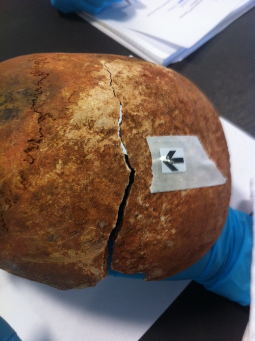
Post-mortem breaks tend to shatter compared to peri-mortem breaks which splinter, this is because bones which are in the post-mortem stage tend to be dry and rather brittle. Another big indicator of a fracture being post-mortem is the difference in colour.

The ‘Quick Tip’ that my applied anthropology lecturer taught me on how to easily distinguish between peri-mortem and post-mortem is to look at the fracture and decide; is it a clean break, as if you were breaking in half a bar of chocolate? If it is, then the fracture is most likely to be a peri-mortem fracture. If the break looks crumbly, like breaking a biscuit in half, it’s post-mortem fracture. Obviously this tip is not the most scientific, but it’s an easy way to begin your distinguishing process.

Image

Skull with signs of post-mortem fractures. This photo is from a practical lab session.

If you look at the photo above it illustrates a post-mortem fracture. You can determine this easily due to the colour difference on the edge of the fracture, where it is a much lighter colour compared to the rest of the skull and the crumbly nature of the cut.

References:

Most of this is my own knowledge that I learnt during my degree in my anthropology lectures/lab practical sessions. But if you’re looking for a published journal check the one below. It is very informative and easy to understand if you’re a beginner in the world of anthropology/archaeology! It also highlights some problems that can arise when distinguishing trauma, it’s really interesting!

Smith, A.C. 2010. Distinguishing Between Antemortem, Perimortem, and Postmortem Trauma. Academia.edu. Available from here in .pdf form!

Read more anthropology/archaeology quick tips here!

Quick Tips: The Use of 3D Animation to Visualise a Crime Scene in Forensics.

Many television programs create 3D animations and computer generated images using highly technical computer programmes to help re-enact the scenes or time frame of a crime. This is mostly used so that the viewer at home can really grasp what crime has been committed and help establish a sense that they are a witness. But in reality these animations and images are becoming an increasingly popular technique used within the courtroom.

Information and evidence can be easily constructed from the traditional methods of forensic photography, blood spatter analysis and eye witness testimonies. But in this modern technological time the information gathered is now being used to create computerised animation that depicts the series of events within a crime. But is this method of providing visual appropriate and correct? Could the animation be showing a display of actions/movements that humans can’t possibly and physically make?

There is a big issue with admissibility, which can cause bias. This occurs when the jurors or judge aren’t aware of an error/uncertainty within the procedure of recreating a real life scene into animation. This can cause them to believe that the evidence is a hundred per cent correct, when in fact there are many errors which were created in the process or animation (Ma & Zheng, 2010). Another big problem arises when studies found that people are five times more likely to remember something they see and hear rather than hearing alone. People are also twice as likely to be persuaded if the arguments are backed with visual evidence (Lederer & Solomon, 1997). So this poses a huge problem as false memories and false testimonies could be influenced, which in the end could cause an innocent person to go to jail for a crime they did not commit.

So with the possibility of creating false memories is the use of 3D animation beneficial for the use of visualising crime scenes within court? It is argued that it is as the use of computerised images creates a higher level of accuracy and speeds up the forensic investigational process but only in major crime types, not every day homicides and robberies. However even though it has limited application in the courtrooms,  it can pose to be very useful in formal briefs with the forensic personnel, and within the backstage elements of the investigation itself (Ma & Zheng, 2010).

References:

Lederer FI, Solomon SH. 1997. Courtroom technology – an introduction to the onrushing future. Fifth National Court Technology Conference: National Centre for State Courts. Available here.

Ma M, Zheng H. 2010. Virtual Reality and 3D Animation in Forensic Visualization. Journal of Forensic Sciences. 55, 5. 1227-1231.

Unusual-ology: How to get A-Head in Medieval England.

 This is a recap of lecture which I attended during my degree. It focused on an extremely weird case study which the lecturer had been presented with, a skull which had preserved soft tissue with ‘links to medieval witchcraft’. 

Within this lecture we were presented with a case study of a skull which had preserved soft tissue. The lecturer talked about how he had stumbled on this rare artefact in a peculiar museum which dealt in Wiccan and witchcraft. There it had a story allocated to it to entice an audience and raise curiosity.

This story was of medieval origins where brutal executions took place. This skull belonging to an execution victim was stored in a Church inside a wooden box covered in numerous markings of figures in communal scenes; these figures are depictions of ‘Green Men’. Intrigued by these mysterious figures and why they appeared on a wooden box along with scrape marks, I decided to do some research of my own. I found that ‘Green Men’ are deities of nature Gods which are often found within Churches and a symbol of pre-Christian religions often in Pagan origin. Could this skull have been kept as a relic? Does it belong to an important figure from medieval times? But after the War was it stolen from the Church and sold onto this museum?

What was peculiar about it was the preserved tissue only found on bodies in extreme conditions such as heat or cold or bodies submerged in bogs. The initial thought was “Could this marvellous story be true? It is a modern day fake, or is it something totally different? Or is it actually a mummy?”

What first stood out was that the skull had remains of eyes which mummies do not possess at the time of burial. Also from examining the skull and comparing the skulls to known parameters it was discovered to be the skull of a 35 to 45 year old woman. More tests were run using modern day technology such as MRI scans and CT scans, the MRI scan failed to show anything due to the lack of hydrogen atoms within the skull due to the dehydrated state it has deteriorated to. But there were surprising results from the CT scan which showed that there was a grey area in the back of the skull, could this be preserved brain? It turned out it was actually tree sap, which backs up the theory that this skull is actually a mummy. Egyptians used pine resin to preserve the bodies as its more ‘sacred’ than glue. There was also evidence that the septum of the skull had been broken by something, which is common in the procedure for mummification. To get a definitive answer to this riddle the final step was to use carbon dating; this helped determine that the skull is in fact a mummy and not a medieval relic or execution victim.

The use of all these modern technologies, theories and knowledge gained from previous case studies helped shine a light on the true origins of this skull. Other artefacts have been found to be something they’re not and are now being traced back to their true beginnings and having a new story assigned to them, for the future generations to be marvelled just as I was during this riveting lecture.

If you’ve enjoyed this new ‘Unsual-ology’ post feature – leave a comment or a like!

Welcome!

Hello and welcome to All Things AAFS!

This blog will discuss and dissect topics of contemporary sciences consisting of journals, published articles or even news that I have stumbled across and wish to expand on.

After finishing my BSc (Hons) degree I have embarked on this task to ensure that I stay up to date with all things AAFS and related to my degree. A few posts on this site will be from lectures or assignments that were set during the course of my degree, which I found fascinating so wish to share with others who are passionate about anything related to archaeological, anthropological and forensic sciences.

Well I hope you enjoy these insights into my experiences in the AAFS world and find them interesting.

Rosie.

Me during DBD'11


Me, inside my designated pit during the DBD’11 Archaeological dig held yearly by Bournemouth University.