Quick Tips: Identifying Dental Diseases – Dental Caries.  

Quick Tips: Identifying Dental Diseases – Dental Caries.  

In our previous Quick Tip post on identifying dental diseases, we gave a basic overview on the disease dental/enamel hypoplasia. If you haven’t read it, you can find it by clicking here.

Dental caries, also known as tooth decay, is thought to be the most common of dental diseases. This is due to it being recorded within archaeological populations more frequently than other dental diseases. It is an infectious and spreadable disease, which is the result of the fermentation of carbohydrates by bacteria that are present within teeth plaque. Its appearance can sometimes be observed as small opaque spots on the crowns of teeth, to large gaping cavities.

dental caries

Dental caries appearance can sometimes be observed as small opaque spots on the crowns of teeth, to large gaping cavities.

Dental caries occurs when sugars from the diet, particularly sucrose, are fermented by the bacteria Lactobacilus acidophilus and Streptococcys mutans, which are found within the built up plaque. This fermentation process causes acids to be produced, which in turn break down and demineralises teeth leaving behind cavities.

Powell (1985) divided the causes of dental caries into different areas, which are;

  • Environmental factors, the trace elements in food and water (i.e fluoride in water sources may protect against caries).
  • Pathogenic factors, the bacterial causing the disease.
  • Exogenous factors, from diet and oral hygiene.
  • Endogenous factors, the shape and structure of teeth.

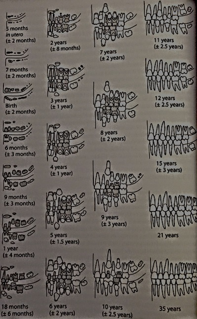
Any part of the tooth structure that allows the accumulation of plaque and food debris can be susceptible to caries. This means that the crowns of the tooth (especially with molars and premolars due to the fissures), and the roots of the teeth are the areas most commonly affected by dental caries.

References:

Lukacs, J.R. 1989. Dental paleopathology: methods for reconstructing dietary patterns. In M.Y. Iscan and K.A.R. Kennedy (eds), Reconstruction of life from the skeleton. New York, Alan Liss, pp. 261-86.

Powell, M.L. 1985. The analysis of dental wear and caries for dietary reconstruction. In R.I. Gilbert and J.H. Mielke (eds), Analysis of prehistoric diets. London, Academic Press, pp. 307-38.

Ubelaker, D.H. 1989. Human Skeletal Remains: Excavation, Analysis, Interpretation (2nd Ed.). Washington, DC: Taraxacum.

White, T.D., Folkens, P.A. 2005. The Human Bone Manual. San Diego, CA: Academic Press. Pg 392-398.

This is the second post of the Quick Tips series on identifying dental diseases. The next post in this series will focus on how to identify calculus (calcified plague), and highlight the cause of this dental disease. To read more Quick Tips in the meantime, click here.

If you’re new to the realm of archaeological, anthropological and forensic sciences (AAFS), or are a student needing sturdy and reliable references, or wondering “what archaeology or anthropology textbooks are good? Check out our new ‘Useful Literature’ page for suggestions from peers and professors!

Quick Tips: Identifying Dental Diseases – Dental/Enamel Hypoplasia.

In our previous Quick Tip post on identifying dental diseases, we gave a basic overview on the different diseases that are observed. If you haven’t read it, you can find it by clicking here.

Dental hypoplasia is a condition that affects the enamel of a tooth. It is characterised by pits, grooves and transverse lines which are visible on the surface of tooth crowns. The lines, grooves and pits that are observed are defects in the enamels development. These defects occur when the enamel formation, also known as amelogenesis, is disturbed by a temporary stress to the organism which upsets the ameloblastic activity. Factors which can cause such stress and therefore disrupt the amelogenesis include; fever, malnutrition, and hypocalcemia.

Figure 1: An example of linear enamel hypoplasia.

Figure 1: An example of linear enamel hypoplasia.

It has been noted that enamel hypoplasia is more regularly seen on anterior teeth than on molars or premolars, and that the middle and cervical portions of enamel crowns tend to show more defects than the incisal third. This is due to the amelogenesis beginning at the occlusal apex of each tooth crown and proceeding rootward, towards where the crown then meets the root at the cervicoenamel line.

Figure 2: Anatomy of a tooth. Note the top third is known as either the occlusal third if in molars, or the incisal third when the tooth is an incisor or canine.

Figure 2: Anatomy of a tooth. Note the top third is known as either the occlusal third if in molars, or the incisal third when the tooth is an incisor or canine.

By studying these incidents of enamel hypoplasia within a population sample, we can be provided with valuable information regarding patterns of dietary stress and disease that may have occurred within the community.

References:

Lukacs, J.R. 1989. Dental paleopathology: methods for reconstructing dietary patterns. In M.Y. Iscan and K.A.R. Kennedy (eds), Reconstruction of life from the skeleton. New York, Alan Liss, pp. 261-86.

Ubelaker, D.H. 1989. Human Skeletal Remains: Excavation, Analysis, Interpretation (2nd Ed.). Washington, DC: Taraxacum.

White, T.D., Folkens, P.A. 2005. The Human Bone Manual. San Diego, CA: Academic Press. Pg 392-398.

This is the second post of the Quick Tips series on identifying dental diseases. The next post in this series will focus on how to identify dental caries and highlight the cause of this dental disease.

To read more Quick Tips in the meantime click here, or to learn about basic fracture types and their characteristics/origins click here!

Quick Tips: Identifying Dental Diseases – The Basics.

Quick Tips: Identifying Dental Diseases – The Basics.

In a previous Quick Tip post we briefly touched on teeth in anthropology/archaeology by providing a basic answer to the question, “What can an anthropologist tell from the examination of teeth?”, which can be found by clicking here.

“No structures of the human body are more likely to disintegrate during life than teeth, yet after death none have greater tenacity against decay” – Wells, 1964.

Teeth are the hardest and most chemically stable tissues in the body; because of this, they’re sometimes the only part of a skeletal remain to withstand the excavation. Even though teeth are the most robust structures of a skeleton, there are numerous diseases that can affect them. This is due to teeth interacting directly with the environment and therefore are vulnerable to damage from physical and biological influences. It is from these diseases, that archaeologists and anthropologists can learn a wealth of information on an individual or population’s diet, oral hygiene, dental care and occupation.

Lukacs, 1989, classified dental diseases into four categories, which are;

  • Infectious – This is one of the more common disease types found within archaeological populations. An example of an infectious dental disease is caries.
  • Degenerative – This is where the dental disease occurs over time as the person ages. An example of degenerative dental disease includes recession of the jaw bone.
  • Developmental –These dental diseases develop due to environmental and lifestyle factors, such as malnutrition. An example of this type of disease is enamel hypoplasia.
  • Genetic – These types of diseases are caused by genetic anomalies.

The main dental diseases that are observed within an archaeological or anthropological context are;

If the dental disease listed above is a link, it means that I have already covered it in an individual blog post and can be found by following the link.

Each of these dental diseases has their own characteristics which allows them to be easily distinguished from one and another. In the next few posts of this Quick Tips series, we will be focusing on each dental disease individually, and highlighting their aetiology and physical characteristics.

References:

Buikstra, J.E., Ubelaker, D.H. 1994. Standards for Data Collection From Human Skeletal Remains. Fayetteville, Arkansas: Arkansas Archaeological Survey Report Number 44.

Lukacs, J.R. 1989. Dental paleopathology: methods for reconstructing dietary patterns. In M.Y. Iscan and K.A.R. Kennedy (eds), Reconstruction of life from the skeleton. New York, Alan Liss. Pg 261-86.

Ubelaker, D.H. 1989. Human Skeletal Remains: Excavation, Analysis, Interpretation (2nd Ed.). Washington, DC: Taraxacum.

Wells, C. 1964. Bones, bodies and disease. London, Thames and Hudson.

White, T.D., Folkens, P.A. 2005. The Human Bone Manual. San Diego, CA: Academic Press. Pg 392-398.

This is the first post of the Quick Tips series on identifying dental diseases. The next post in this series will focus on how to identify dental/enamel hypoplasia and highlight the cause of this dental disease.

To read more Quick Tips in the meantime, click here, or to learn about basic fracture types and their characteristics/origins click here!

Quick Tips – Common Questions: What can an anthropologist tell from the examination of teeth regarding either forensic identification of individuals or understanding past populations?

This is a Quick Tips post providing a basic answer to a commonly asked question often faced within the field of archaeology and anthropology.

An anthropologist can obtain a wide and varied collection of information from examining teeth. Information such as paleodiets and palaeoenvironments can be learnt from studying a population, or from studying an individual sample you can identify how old the person was at time of death or whether that person was pregnant/ill. These examples are just the tip of the iceberg on what you can learn from dentition.

Ondontology

An anthropologist can obtain a wide and varied collection of information from examining teeth, ranging from palaeodiets and palaeoenvironmental information to age of death.

From studying a large population dentition sample, a picture can be painted of their past diets, current diets and palaeoenvironments. Isotopes play a huge part in conducting research into palaeodiets and palaeoenvironments.

Isotopes are deposited into the teeth of an individual/population from food sources or environment. A tooth can provide isotopic information from the past 20yrs of the individual’s life. The enamel and dentine can be examined to analyse the isotopic values that will pinpoint an origin of a population or food sources. The carbon and nitrogen isotope compositions found within the enamel are used to reconstruct diet and the oxygen isotopes are used to determine the geographic origin of the food source. The carbon isotopes are absorbed from the diet of the animals that are sources and the oxygen isotopes from the water that the population consume. These isotopic values are vital in helping an anthropologist understand the local ecosystem a population exploited and whether a population migrated to numerous locations which caused changes in the available diet.

The cementum of a tooth can highlight important information about a person which can be used for forensic identification; this information could give an approximate age of death. An example of this application is seen in Kagerer and Grupe (2000) study where they obtained 80 freshly extracted teeth and investigated the incremental lines in acellular extrinsic fibre cementum. From studying the cementum, they were able to determine the age of the patient by comparing it to detailed queries of the patients life history. This study also identified patients who were pregnant. Kagerer and Grupe (2000) concluded that if there was a presence of hypo-mineralised incremental lines on the extracted tooth, the patient was pregnant. This is due to the pregnancies influence on calcium metabolism. A confliction with this is that hypo-mineralized lines can also appear when a skeletal trauma or renal illness was present.

By looking at the dentition of molars the age of the skeleton can be estimated. A recent study by Mesotten, et al. (2002) highlighted the application of forensic odontology. Mesotten, et al’s methodology consisted of examining 1175 orthopantomograms which belonged to patients who were of Caucasian origin and were aged between 16 and 22years. From their investigation Mesotten, et al. were able to conclude that from studying the molars, it was possible to age Caucasian individuals with a regression formula with a standard deviation of 1.52 or 1.56 years for males and females, respectively, if all four third molars were available. This could play a fundamental role in identifying a missing person by estimating the decease’s age and seeing if its estimate matches the individual.

Although the studies from Mesotten, et al (2002) and Kagerer and Grupe (2000) have been written about and applied to individual cases, their methodology and conclusions can be applied to a past population if a group of skeletons were found with preserved teeth. The individual’s age of death can be used as quantitative data, alongside other individuals from the same sample, to figure out a past population’s life expectancy.

References:

Kagerer, P. Grupe, G. 2000. Age-at-death diagnosis and determination of life-history parameters by incremental lines in human dental cementum as an identification aid. Forensic Science International. 118, 1. 75-82.

Mesotten, K. Gunst, K. Carbonez, A. Willems, G. 2002. Dental age estimation and third molars: a preliminary study. Forensic Science International. Volume 129, Issue 2, 110-115

To learn how archaeologists and anthropologists use teeth to age skeletal remains, read our Quick Tips: How To Estimate The Chronological Age of a Human Skeleton – Using Dentition to Age Subadults. Or to read more of our interesting Quick Tips, click here.

Quick Tips: How To Estimate The Chronological Age Of A Human Skeleton – Using Dentition To Age Subadults.

This Quick Tips post is the third in the series on age estimation of human skeletal remains, if you haven’t read the first post click here to start at the beginning. The first post provides an overview of the different techniques utilised by archaeologists/anthropologists, which will each be covered in more detail in their own blog post, and the categories that human skeletal remains are placed under according to their chronological age. The second post examines the epiphyseal closure method, which you can find here.

The practice of using dentition to chronologically age human skeletal remains is split into two halves, depending on the whether the skeleton is that of a subadult or adult. This blog post is going to discuss using dentition to age subadults.

Due to the abundance of teeth found in many archaeological, forensic, paleontological, and anthropological contexts and because of the regular tooth formation and eruption times, dental development is the most widely used technique for aging subadult remains. As stated in my previous blog post, several elements of the human skeleton begin the stages of epiphyseal fusion alongside the conclusion of tooth eruption; these two techniques (dentition and epiphyseal closure) are often used complementary to each other to help age sub-adults. When it comes to subadult tooth emergence there are four stages:

Stage 1 is where most of the deciduous teeth, commonly referred to as ‘milk teeth’, emerge during the second year of life.

Stage 2, during this stage the two permanent incisors and the first permanent molar emerge, this stage typically occurs between the age of six and eight years.

Stage 3, occurs between the age of ten and twelve and it involves the emergence of the permanent canines, premolars, and second molars.

Stage 4, or the final stage involves the third molar emerging around the age of eighteen years.

When looking at dentition you must look at all aspects of emergence and not just at the fully erupted tooth, which includes the completeness of all roots and crowns (formation) and the position of each tooth relative to the alveolar margin (eruption). Ubelaker (1989) conducted a study on non-Native Americans and created a graphic summary of dental development and the correlating ages it occurs, see figure 1.

Figure 1: Ubelaker's (1989) diagram showing the dental development in correlation to age.

Figure 1: Ubelaker’s (1989) diagram showing the dental development in correlation to age.

It must be noted that the ages these stages occur at differ per individual so only act as a reference. Gustafson and Koch (1974) created a graph to illustrate the variation that could occur with dental development, see figure 2.

Figure 2: Gustafson and Koch's (1974) image showing the variation in timing of dental development. Colour key: Black highlights the age that crown mineralization begins, Dark grey shows the age of crown completion, Light grey shows the age of eruption, and White displays age of root completion.

Figure 2: Gustafson and Koch’s (1974) image showing the variation in timing of dental development. Colour key: Black highlights the age that crown mineralization begins, Dark grey shows the age of crown completion, Light grey shows the age of eruption, and White displays age of root completion.

References:

Gustafson, G. Koch, G. 1974. Age estimation up to 16 years of age based on dental development. Odontologisk Revy. 25. Pg 297-306.

Ubelaker, D.H. 1989. Human Skeletal Remains: Excavation, Analysis, Interpretation (2nd Ed.). Washington, DC: Taraxacum.

White, T.D., Folkens, P.A. 2005. The Human Bone Manual. San Diego, CA: Academic Press. Pg 360-385.

This is the third of a Quick Tips series on ageing skeletal remains, the next in this series will focus on the use of dentition to age adults and the use of cranial suture closure. To read more Quick Tips in the meantime, click here

To learn about basic fracture types and their characteristics/origins in their own Quick Tips series, click here!