Quick Tips: How To Estimate The Chronological Age Of A Human Skeleton – Sternal Rib End Method.

This Quick Tips post is the sixth in the series on age estimation on skeletal remains, if you haven’t read the previous post click here, or to start at the beginning click here. The previous post provides an overview of the pubic symphyseal surface method of ageing, whereas the first post covers the basics.

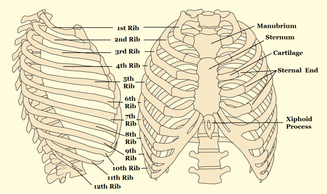
The method was primarily developed by Iscan and Loth (1986) who studied the metamorphosis of the sternal end of the fourth rib. They found that the metamorphosis corresponds to the age but does vary by sex.

In their study they examined the “form, shape, texture and overall quality” of the sternal end which is found at the anterior (ventral) end of the shaft. This end is a roughened, porous, cupped oval surface which attaches to the cartilage attached to the sternum.  From this they were able to define a series of phases that depict the metamorphism of the sternal rib end over time.

Rib anatomy

Anatomy of the rib cage. This method was primarily developed by Iscan and Loth (1986) who studied the metamorphosis of the sternal end of the fourth rib. They found that the metamorphosis corresponds to the age but does vary by sex.

At the start the sternal end is flat or billowy with regular and rounded edges, and over time its rim thins and become irregular, with the surface porosity increasing, and the end becomes irregular. This method can be applied cautiously to the 3rd or 5th ribs as well, but not the others.

References:

Iscan, M.Y., and Loth, S.R. 1986. Estimation of age and determination of sex from the sternal rib. In: K. J. Reichs (ed.) Forensic Osteology: Advances in the Identification of Human Remains. Springfield, Illinois. Pg 68-89.

White, T.D., Folkens, P.A. 2005. The Human Bone Manual. San Diego, CA: Academic Press. Pg 360-385.

If you’re new to the realm of archaeological, anthropological and forensic sciences (AAFS), or are a student needing sturdy and reliable references, or wondering “what archaeology or anthropology textbooks to buy? Check out our new ‘Useful Literature’ page!


Quick Tips: Archaeological Techniques –Use of Isotopes in Archaeology.

Isotopic analysis is widely used within the worlds of archaeology and anthropology. From analysing isotopes we’re able to uncover a wide range of information regarding the past; ranging from palaeoenvironments to palaeodiets, and even using isotopes to reconstruct trade routes of materials.

But first, what are isotopes?

All of the chemical elements consist of atoms which are specific to the element and the mass of an atom is dictated by the number of protons and neutrons it contains. The identity of the chemical element depends on the number of protons found within the atom’s nucleus, but the number of neutrons within the atom can vary. Atoms of the same chemical element (same number of protons), but with different masses, which is from the varying amount of neutrons, are called isotopes.

Stone Circle at Drombeg

Within nature, most of the elements consist of a number of isotopes. These isotopes can be found within water, livestock, crops and plants, which can then be used to reconstruct palaeodiets and palaeoenvironments.

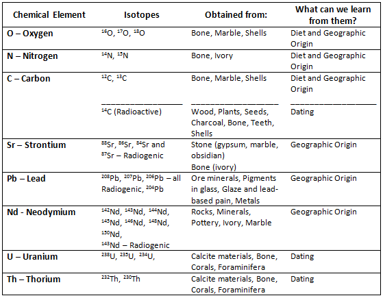
Within nature, most of the elements consist of a number of isotopes. For a great majority of elements these relative proportions of isotopes are fixed, but there are a group of elements which either due to chemical or biochemical processes are of variable isotopic composition. These elements are oxygen, carbon, nitrogen and sulphur. Another group of isotopes that are used for analysis are strontium, lead and neodymium. These are formed by elements which contain stable but radiogenic isotopes, which are formed by radioactive decay of another element. Carbon and nitrogen isotope composition are primarily used to reconstruct diets, and oxygen isotopes are used to determine geographic origin. Strontium and lead isotopes found within teeth and bone can sometimes be used to reconstruct migration patterns in human populations and cultural affinity

Isotopes Table

A table of the various elemental isotopes that are valuable in archaeological and anthropological research.

But how do isotopes get into skeletal remains?

Carbon isotopes are taken up through the diet of animals during their lifetime and these isotopes are deposited into teeth and bones of humans when they are consumed and digested. By studying animal bones and examining the 12C and 13C isotope ratio, it is possible to determine whether the animals ate predominately 3C or 4C plants. Oxygen isotopes are constantly being taken up and deposited into the body through the water a population drinks. This process ends with the organism’s death, from this point on isotopes no longer accumulate in the body, but do undergo degradation. For best result the researcher would need to know the original levels, or estimation thereof, of isotopes in the organism at the time of its death.

By creating a map of these natural occurring isotopes in different environments, rivers and areas, it is possible to identify where in an area the population lived, sourced their water or where the livestock grazed, by comparing the levels of isotopes that were obtained from skeletal remains to the environmental map. This mapping can also help identify trade routes that once existed and can also identify the migration patterns of populations.

References:

Balme, J., Paterson, A. 2006. Archaeology in Practice: A Student Guide to Archaeological Analayses. Oxford, UK: Blackwell Publishing. Pg 218.

Renfrew, C., Bahn, P. 1991. Archaeology: Theories, Methods and Practice. London, UK: Thames & Hudson. Pg 249-53.

If you’re new to the realm of archaeological, anthropological and forensic sciences (AAFS), or are a student needing sturdy and reliable references, or wondering “what archaeology or anthropology textbooks to buy? Check out our new ‘Useful Literature’ page!

Quick Tips – Common Questions: Why are some diseases more easily identified on skeletal remains than others?

This is a Quick Tips post providing a basic answer to a commonly asked question often faced within the field of archaeology and anthropology.

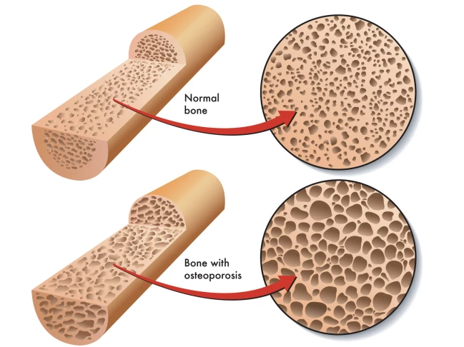
Some diseases are more easily to identify on skeletal remains due to leaving tell-tale signs in the bones preservation. An easy example of this is osteoporosis; this condition leaves the inners of bones a lot more porous which is easier to visually assess and compare to a ‘healthy’ individual’s skeletal remains.

canstockphoto18602582

Some diseases are more easily to identify on skeletal remains due to leaving tell-tale signs in the bones preservation. An  example of this is osteoporosis; this condition leaves the inners of bones a lot more porous than normal bones.

A study by Hershkovitz & Rothschild (1997) highlighted how certain medical conditions, in their study sickle cell anaemia, affects the bone growth and development. Hershkovitz & Rothschild found that due to the iron deficiency from sickle cell anaemia caused porotic hyperostosis (symmetrical osteoporosis) on the parietal bone as well as others. They were able to visually diagnose this due to the characteristic ‘pores’ over the skull.

Another example of an easily identifiable disease is tuberculosis (TB), TB can cause devastating bone damage. A recent archaeological study by Lewis (2011) looked into a population who suffered from TB. Lewis visually analysed the skeletal remains of a juvenile population from Poundbury Camp, Dorset. The TB infection caused numerous ailments to the infected, such as fever, but it’s the skeletal damage which gave the indication that the person suffered.  Amongst the population there was a high instance of skeletons with necrosis and lytic lesions characterised by minimal bone formation.  Many of the juvenile’s vertebrae displayed new bone formations which could indicate the presence of a paravertebral abscess. Many of the metatarsals were also displaying evidence of new bone formation which they concluded could be indicative of tuberculous dactylitis. Osteomyelitis, infection of the bone, was also found on a few mandibles and visually diagnosed due to its characteristic small pores found in a localised area. It is this characterised skeletal damage, seen on numerous cases during known TB outbreaks, which cause more diseases to be easily identified by eye due to the skeletal anomalies.

There are problems when trying to differentiate certain diseases for example; TB with brucellosis (undulant fever). As they both produce spinal lesions it is necessary to observe the other characteristic skeletal damage (new bone formation and osteomyelitis) to correctly identify it as a TB infection. Another slight difference between TB and brucellosis is that the spinal lesions are more sclerotic and regular than those from a TB infection (Lewis, 2011).

These porous bones and unexpected bone formations are easily observed, as they are not what’s expected during the known skeletal development found in healthy persons. Problems arise with diseases that do no damage to the skeleton, but instead affect soft tissue and muscles. These illnesses are harder to identify as they decay over time leaving only trace elements in the surrounding soils which would then hold the key for disease identification.

References:

Hershkovitz, I. Rothschild, B. et al. 1997. Recognition of sickle cell anemia in skeletal remains of children. American Journal of Physical Anthropology. Volume 104, Issue 2. 213-226.

Lewis, M. 2011. Tuberculosis in the non-adults from Romano-British Poundbury Camp, Dorset, England. International Journal of Paleopathology. Volume 1, Issue 1. 12-23.

To learn how archaeologists and anthropologists use teeth to age skeletal remains, read our Quick Tips: How To Estimate The Chronological Age of a Human Skeleton – Using Dentition to Age Subadults. Or to read more of our interesting Quick Tips, click here.

Quick Tips: Identifying Dental Diseases – Dental Caries.  

Quick Tips: Identifying Dental Diseases – Dental Caries.  

In our previous Quick Tip post on identifying dental diseases, we gave a basic overview on the disease dental/enamel hypoplasia. If you haven’t read it, you can find it by clicking here.

Dental caries, also known as tooth decay, is thought to be the most common of dental diseases. This is due to it being recorded within archaeological populations more frequently than other dental diseases. It is an infectious and spreadable disease, which is the result of the fermentation of carbohydrates by bacteria that are present within teeth plaque. Its appearance can sometimes be observed as small opaque spots on the crowns of teeth, to large gaping cavities.

dental caries

Dental caries appearance can sometimes be observed as small opaque spots on the crowns of teeth, to large gaping cavities.

Dental caries occurs when sugars from the diet, particularly sucrose, are fermented by the bacteria Lactobacilus acidophilus and Streptococcys mutans, which are found within the built up plaque. This fermentation process causes acids to be produced, which in turn break down and demineralises teeth leaving behind cavities.

Powell (1985) divided the causes of dental caries into different areas, which are;

  • Environmental factors, the trace elements in food and water (i.e fluoride in water sources may protect against caries).
  • Pathogenic factors, the bacterial causing the disease.
  • Exogenous factors, from diet and oral hygiene.
  • Endogenous factors, the shape and structure of teeth.

Any part of the tooth structure that allows the accumulation of plaque and food debris can be susceptible to caries. This means that the crowns of the tooth (especially with molars and premolars due to the fissures), and the roots of the teeth are the areas most commonly affected by dental caries.

References:

Lukacs, J.R. 1989. Dental paleopathology: methods for reconstructing dietary patterns. In M.Y. Iscan and K.A.R. Kennedy (eds), Reconstruction of life from the skeleton. New York, Alan Liss, pp. 261-86.

Powell, M.L. 1985. The analysis of dental wear and caries for dietary reconstruction. In R.I. Gilbert and J.H. Mielke (eds), Analysis of prehistoric diets. London, Academic Press, pp. 307-38.

Ubelaker, D.H. 1989. Human Skeletal Remains: Excavation, Analysis, Interpretation (2nd Ed.). Washington, DC: Taraxacum.

White, T.D., Folkens, P.A. 2005. The Human Bone Manual. San Diego, CA: Academic Press. Pg 392-398.

This is the second post of the Quick Tips series on identifying dental diseases. The next post in this series will focus on how to identify calculus (calcified plague), and highlight the cause of this dental disease. To read more Quick Tips in the meantime, click here.

If you’re new to the realm of archaeological, anthropological and forensic sciences (AAFS), or are a student needing sturdy and reliable references, or wondering “what archaeology or anthropology textbooks are good? Check out our new ‘Useful Literature’ page for suggestions from peers and professors!

Quick Tips: Identifying Dental Diseases – Dental/Enamel Hypoplasia.

In our previous Quick Tip post on identifying dental diseases, we gave a basic overview on the different diseases that are observed. If you haven’t read it, you can find it by clicking here.

Dental hypoplasia is a condition that affects the enamel of a tooth. It is characterised by pits, grooves and transverse lines which are visible on the surface of tooth crowns. The lines, grooves and pits that are observed are defects in the enamels development. These defects occur when the enamel formation, also known as amelogenesis, is disturbed by a temporary stress to the organism which upsets the ameloblastic activity. Factors which can cause such stress and therefore disrupt the amelogenesis include; fever, malnutrition, and hypocalcemia.

Figure 1: An example of linear enamel hypoplasia.

Figure 1: An example of linear enamel hypoplasia.

It has been noted that enamel hypoplasia is more regularly seen on anterior teeth than on molars or premolars, and that the middle and cervical portions of enamel crowns tend to show more defects than the incisal third. This is due to the amelogenesis beginning at the occlusal apex of each tooth crown and proceeding rootward, towards where the crown then meets the root at the cervicoenamel line.

Figure 2: Anatomy of a tooth. Note the top third is known as either the occlusal third if in molars, or the incisal third when the tooth is an incisor or canine.

Figure 2: Anatomy of a tooth. Note the top third is known as either the occlusal third if in molars, or the incisal third when the tooth is an incisor or canine.

By studying these incidents of enamel hypoplasia within a population sample, we can be provided with valuable information regarding patterns of dietary stress and disease that may have occurred within the community.

References:

Lukacs, J.R. 1989. Dental paleopathology: methods for reconstructing dietary patterns. In M.Y. Iscan and K.A.R. Kennedy (eds), Reconstruction of life from the skeleton. New York, Alan Liss, pp. 261-86.

Ubelaker, D.H. 1989. Human Skeletal Remains: Excavation, Analysis, Interpretation (2nd Ed.). Washington, DC: Taraxacum.

White, T.D., Folkens, P.A. 2005. The Human Bone Manual. San Diego, CA: Academic Press. Pg 392-398.

This is the second post of the Quick Tips series on identifying dental diseases. The next post in this series will focus on how to identify dental caries and highlight the cause of this dental disease.

To read more Quick Tips in the meantime click here, or to learn about basic fracture types and their characteristics/origins click here!

Quick Tips: Archaeological Techniques – Aerial Photography.

Aerial photography is a surveying technique that involves taking a photographic record from satellites, aircrafts and balloons, to aid with the detection of buried archaeological remains and features, which may be difficult to identify at ground level.

There are two types of aerial photography;

  • Oblique – Oblique aerial photography involves taking a photograph from lower altitudes at an angle. This gives a better perspective and a pictorial effect, and allows for identification of earthworks.
  • Vertical – Vertical aerial photography involves taking a photograph from great heights directly above an area. This gives a bird’s eye view of an area, allowing for easier map making and identification of crop marks.
aerialphoto

Fig. 1: There are two types of aerial photography; oblique and vertical.

These aerial photographs can show numerous phenomena, some of which are sometimes not from archaeological origins. These phenomena include:

  • Crop marks – These types of marks develop when a buried wall or ditch increases or decreases crop growth; this is due to the feature affecting the availability of moisture and nutrients in the soil.
crop mark

Fig 2. An example of a crop mark. You can see in the excavation site the ditch that is affecting the crop’s growth.

  • Soil marks – These marks are caused by changes in the subsoil colour, when a plough brings part of the buried feature to the surface.
Fig. 3: After this field was ploughed, it has exposed the feature which has had parts brought to the surface.

Fig. 3: An example of soil marks. After this field had been ploughed, this buried feature had parts brought to the surface which has caused discolouration in the soil.

  • Earthworks – This phrase is used to describe any features seen in relief. These are also known as shadow marks when viewed from the air.
Fig. 4: This is an example of an earthwork. This particular archaeological site is an abandoned Medieval settlement.

Fig. 4: This is an example of an earthwork. This particular archaeological site is an abandoned Medieval settlement.

It is from these phenomena that we’re able to identify whether there is buried archaeology in an area which can then allow for an in-depth investigation.

References:

Balme, J., Paterson, A. 2006. Archaeology in Practice: A Student Guide to Archaeological Analayses. Oxford, UK: Blackwell Publishing. Pg 218.

Renfrew, C., Bahn, P. 1991. Archaeology: Theories, Methods and Practice. London, UK: Thames & Hudson. Pg 249-53.

Click here to read more Quick Tip posts!


Quick Tips: Identifying Dental Diseases – The Basics.

Quick Tips: Identifying Dental Diseases – The Basics.

In a previous Quick Tip post we briefly touched on teeth in anthropology/archaeology by providing a basic answer to the question, “What can an anthropologist tell from the examination of teeth?”, which can be found by clicking here.

“No structures of the human body are more likely to disintegrate during life than teeth, yet after death none have greater tenacity against decay” – Wells, 1964.

Teeth are the hardest and most chemically stable tissues in the body; because of this, they’re sometimes the only part of a skeletal remain to withstand the excavation. Even though teeth are the most robust structures of a skeleton, there are numerous diseases that can affect them. This is due to teeth interacting directly with the environment and therefore are vulnerable to damage from physical and biological influences. It is from these diseases, that archaeologists and anthropologists can learn a wealth of information on an individual or population’s diet, oral hygiene, dental care and occupation.

Lukacs, 1989, classified dental diseases into four categories, which are;

  • Infectious – This is one of the more common disease types found within archaeological populations. An example of an infectious dental disease is caries.
  • Degenerative – This is where the dental disease occurs over time as the person ages. An example of degenerative dental disease includes recession of the jaw bone.
  • Developmental –These dental diseases develop due to environmental and lifestyle factors, such as malnutrition. An example of this type of disease is enamel hypoplasia.
  • Genetic – These types of diseases are caused by genetic anomalies.

The main dental diseases that are observed within an archaeological or anthropological context are;

If the dental disease listed above is a link, it means that I have already covered it in an individual blog post and can be found by following the link.

Each of these dental diseases has their own characteristics which allows them to be easily distinguished from one and another. In the next few posts of this Quick Tips series, we will be focusing on each dental disease individually, and highlighting their aetiology and physical characteristics.

References:

Buikstra, J.E., Ubelaker, D.H. 1994. Standards for Data Collection From Human Skeletal Remains. Fayetteville, Arkansas: Arkansas Archaeological Survey Report Number 44.

Lukacs, J.R. 1989. Dental paleopathology: methods for reconstructing dietary patterns. In M.Y. Iscan and K.A.R. Kennedy (eds), Reconstruction of life from the skeleton. New York, Alan Liss. Pg 261-86.

Ubelaker, D.H. 1989. Human Skeletal Remains: Excavation, Analysis, Interpretation (2nd Ed.). Washington, DC: Taraxacum.

Wells, C. 1964. Bones, bodies and disease. London, Thames and Hudson.

White, T.D., Folkens, P.A. 2005. The Human Bone Manual. San Diego, CA: Academic Press. Pg 392-398.

This is the first post of the Quick Tips series on identifying dental diseases. The next post in this series will focus on how to identify dental/enamel hypoplasia and highlight the cause of this dental disease.

To read more Quick Tips in the meantime, click here, or to learn about basic fracture types and their characteristics/origins click here!

Quick Tips: How to Estimate the Biological Sex of a Human Skeleton – Pelvic Dimorphism.

This is the 3rd blog post in this Quick Tips series on estimating the biological sex of human skeletal remains. If you haven’t read the first post on the basics of sexing skeletal remains, click here to start at the beginning or if you skipped the 2nd post focusing on the skull method if sex estimation, click here.

When it comes to sexing skeletal remains by the pelvic elements there are a few trends, as stated in the first blog post in this series, the female pelvic bones, specifically the sacra and ossa coxa are smaller and less robust than their male counterparts.

Figure 1: Side by side size comparison of a male (left) and female (right) pelvis.

Figure 1: Side by side size comparison of a male (left) and female (right) pelvis.

Although the female pelvic components are smaller in general, many aspects of the female pelvis are wider than males. The pelvic inlets on a female are relatively wider than those of males, as well as the greater sciatic notches – which is thought to aid childbirth.

Figure 2: Basic annotated diagram of the pelvis.

Figure 2: Basic labelled diagram of the pelvic anatomy.

There are numerous features of the pelvic bones that are examined to identify the biological sex of an individual, alongside the trends stated about. These features are as follows;

  • The ventral arc.
  • The subpubic concavity.
  • The medial aspect of the ischiopubic ramus.
  • The greater sciatic notch.

The first three features listed above, are known as the Phenice method – which was proposed by T. W. Phenice in 1969. His paper, “A Newly Developed Visual Method of Sexing the Os Pubis”, contributed greatly to the method of visual determination of sex, as beforehand the methods were subjective and based largely on the osteologist’s experience. The Phenice method should only be used for fully adult skeletal remains, where it is 96 to 100% accurate.

The ventral arc is a slightly raised ridge of bone that sweeps inferiorly and laterally across the central surface of the pubis. It joins with the medial border of the ischiopubic ramus. The ventral arc is only present in females, although males may have raised ridges in this area, but these do not take the wide, evenly arching appearance of the ventral arc.

Figure 2: The ventral arc is characterised by a slightly raised ridge of bone. Males do not exhibit the ventral arc, where as females do.

Figure 3: The ventral arc is characterised by a slightly raised ridge of bone. Males (left) do not exhibit the ventral arc, where as females (right) do.

To observe the subpubic concavity, you should turn the pubis so that the convex dorsal surface if facing you. Then you should view the medial edge of the ischiopubic ramus. Females display a subpubic concavity here where the edge of the ramus is concaved, whereas males tend to have straight edges or very slightly concaved.
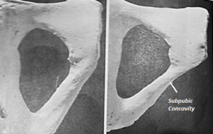
Figure 4: Females display a subpubic concavity here where the edge of the ramus is concaved, whereas males tend to have straight edges or very slightly concaved.

Figure 4: Females (right) display a subpubic concavity here where the edge of the ramus is concaved, whereas males (left) tend to have straight edges or very slightly concaved.

To observe the medial aspect of the ischiopubic ramus, you should turn the pubis 90° so that the symphyseal surface is directly facing you. View the part of the ramus that is directly inferior to the pubis symphysis. In females, the ramus has a sharp, narrow edge, whereas in males it is flat and blunt.

Figure 5: In females (right), the medial aspect of the ischiopubic ramus has a sharp, narrow edge, whereas in males (left) it is flat and blunt.

Figure 5: In females (right), the medial aspect of the ischiopubic ramus has a sharp, narrow edge, whereas in males (left) it is flat and blunt.

As with the five features of the skull used to sex a skeleton in the previous, the greater sciatic notch has also been given a numerical score from 1 to 5 relating to the level of expression. It has been generally found that female os coxae are more likely to exhibit a lower level of expression, whereas male os coxae are more likely to have higher levels of expression.

Figure 6:

Figure 6: It has been generally found that female os coxae are more likely to exhibit a lower level of expression, whereas male os coxae are more likely to have higher levels of expression, when it comes to the greater sciatic notch.

To obtain the best results whist examining the os coxae, it should be held in the same orientation as the pictured above. This allows you to match the angle of the greater sciatic to the closest expression that represents it. It should be noted that this method is usually used as a secondary indicator.

References:

Buikstra, J.E., Ubelaker, D.H. 1994. Standards for Data Collection From Human Skeletal Remains. Fayetteville, Arkansas: Arkansas Archaeological Survey Report Number 44.

Ubelaker, D.H. 1989. Human Skeletal Remains: Excavation, Analysis, Interpretation (2nd Ed.). Washington, DC: Taraxacum.

White, T.D., Folkens, P.A. 2005. The Human Bone Manual. San Diego, CA: Academic Press. Pg 392-398.

This is the third post of the Quick Tips series on sex determination of skeletal remains. The next post in this series will focus on the use of DNA to determine biological sex. To read more Quick Tips in the meantime, click here

Quick Tips – Common Questions: What can an anthropologist tell from the examination of teeth regarding either forensic identification of individuals or understanding past populations?

This is a Quick Tips post providing a basic answer to a commonly asked question often faced within the field of archaeology and anthropology.

An anthropologist can obtain a wide and varied collection of information from examining teeth. Information such as paleodiets and palaeoenvironments can be learnt from studying a population, or from studying an individual sample you can identify how old the person was at time of death or whether that person was pregnant/ill. These examples are just the tip of the iceberg on what you can learn from dentition.

Ondontology

An anthropologist can obtain a wide and varied collection of information from examining teeth, ranging from palaeodiets and palaeoenvironmental information to age of death.

From studying a large population dentition sample, a picture can be painted of their past diets, current diets and palaeoenvironments. Isotopes play a huge part in conducting research into palaeodiets and palaeoenvironments.

Isotopes are deposited into the teeth of an individual/population from food sources or environment. A tooth can provide isotopic information from the past 20yrs of the individual’s life. The enamel and dentine can be examined to analyse the isotopic values that will pinpoint an origin of a population or food sources. The carbon and nitrogen isotope compositions found within the enamel are used to reconstruct diet and the oxygen isotopes are used to determine the geographic origin of the food source. The carbon isotopes are absorbed from the diet of the animals that are sources and the oxygen isotopes from the water that the population consume. These isotopic values are vital in helping an anthropologist understand the local ecosystem a population exploited and whether a population migrated to numerous locations which caused changes in the available diet.

The cementum of a tooth can highlight important information about a person which can be used for forensic identification; this information could give an approximate age of death. An example of this application is seen in Kagerer and Grupe (2000) study where they obtained 80 freshly extracted teeth and investigated the incremental lines in acellular extrinsic fibre cementum. From studying the cementum, they were able to determine the age of the patient by comparing it to detailed queries of the patients life history. This study also identified patients who were pregnant. Kagerer and Grupe (2000) concluded that if there was a presence of hypo-mineralised incremental lines on the extracted tooth, the patient was pregnant. This is due to the pregnancies influence on calcium metabolism. A confliction with this is that hypo-mineralized lines can also appear when a skeletal trauma or renal illness was present.

By looking at the dentition of molars the age of the skeleton can be estimated. A recent study by Mesotten, et al. (2002) highlighted the application of forensic odontology. Mesotten, et al’s methodology consisted of examining 1175 orthopantomograms which belonged to patients who were of Caucasian origin and were aged between 16 and 22years. From their investigation Mesotten, et al. were able to conclude that from studying the molars, it was possible to age Caucasian individuals with a regression formula with a standard deviation of 1.52 or 1.56 years for males and females, respectively, if all four third molars were available. This could play a fundamental role in identifying a missing person by estimating the decease’s age and seeing if its estimate matches the individual.

Although the studies from Mesotten, et al (2002) and Kagerer and Grupe (2000) have been written about and applied to individual cases, their methodology and conclusions can be applied to a past population if a group of skeletons were found with preserved teeth. The individual’s age of death can be used as quantitative data, alongside other individuals from the same sample, to figure out a past population’s life expectancy.

References:

Kagerer, P. Grupe, G. 2000. Age-at-death diagnosis and determination of life-history parameters by incremental lines in human dental cementum as an identification aid. Forensic Science International. 118, 1. 75-82.

Mesotten, K. Gunst, K. Carbonez, A. Willems, G. 2002. Dental age estimation and third molars: a preliminary study. Forensic Science International. Volume 129, Issue 2, 110-115

To learn how archaeologists and anthropologists use teeth to age skeletal remains, read our Quick Tips: How To Estimate The Chronological Age of a Human Skeleton – Using Dentition to Age Subadults. Or to read more of our interesting Quick Tips, click here.

Quick Tips – Common Questions: Can physical activities undertaken during life be detected on skeletal remains?

This is a Quick Tips post providing a basic answer to a commonly asked question often faced within the field of archaeology and anthropology.

Can physical activities undertaken during life be detected on skeletal remains? Yes they can.

Numerous activities, such as hunting, gathering, exercise and more obviously fighting, can inflict damage or adaptations onto to a skeletal system. Some physical activities can be easily identified by due to the damage they can produce to the skeleton, i.e. fighting, whereas the skeletons adapt to strain caused by sport or a daily activity can be harder to detect.

Stock (2006) investigated hunter-gatherer postcranial robusticity relative to patterns of mobility and climatic adaption. In this study, Stock took four collections of known hunter-gatherers skeletal remains along with the associated data of the environmental factors in the population area and the terrestrial mobility. In every analysis conducted, the effective environmental temperature was found to be negatively correlated with strength. Stock concluded that hunter-gatherers from colder climates tend to have stronger long bone diaphysis, than the groups from warmer regions. Although in contrast, the partial correlations between mobility and robusticity are positive; suggesting that activity has a consistently positive relationship with diaphyseal strength. This study indicates that even the simple ‘easy’ activity of hunting and gathering can affect diaphyseal strength of a skeleton and that the activity can be detected.

Exercise is also one of the most common factors to cause a skeleton to adapt. A recent study by Shaw (2009) was able to correctly predict an athlete’s chosen sport from quantifying the soft tissue properties and bone morphology. In Shaw’s study he focused on examining modern athletes (runners, field hockey players, swimmers, and cricketers) and a control group. Using peripheral quantitative computed tomography (pQCT), Shaw quantified the relationship between the amount of muscle and other soft tissues and the morphology of the bones along the midshaft of the arm, forearm and lower legs. This study concluded that Shaw could correctly identify an athlete’s chosen sport from examining a skeletal system and quantifying the bone mass and strength. Shaw concluded that the changes to the bones structural properties were from the strain of daily habitual training from the athlete’s young age.

These two modern studies, Stock (2006) and Shaw (2009), perfectly highlight how physical activities can be detected on skeletal remains.  But these morphological changes can be harder to detect than more brutal activities such as fighting. This is because war and fights leave tell-tale marks on the skeletons which are detectable from eye rather than quantitating data. Violence within a population whether its ritual/habitual, in times of war or domestic can be easily identified from the fractures and dents a bone receives.

A recent NAI (Non-accidental Injury) study from Day et al (2006), highlighted how skeletal remains could indicate bone trauma caused by violence. The study retrospectively observed cases of suspected NAI injuries sustained by children from X-rays obtained at an Edinburgh hospital. The bone fractures, mostly found on the skull and long bones, were suspected to be cause by domestic abuse and evidence of blunt force trauma was observed in numerous cases. Even though this is a recent study conducted on NAI instances, it does appropriately show how violence can inflict damage onto skeletal remains. An archaeological skeleton could show healed/unhealed fractures sustained via a physically demanding activity which was violent in nature, such as war or ritual fighting.

References:

Day, F. Clegg, S. McPhillips, M. Mok, J. 2006. A retrospective case series of skeletal surveys in children with suspected non-accidental injury. Journal of Clinical Forensic Medicine. 13, 12. 55-59.

Shaw, C. 2009. ‘Putting flesh back onto the bones?’ Can we predict soft tissue properties from skeletal and fossil remains?. Journal of Human Evolution. 59, 5. 484-492.

Stock, J.T. 2006. Hunter-Gatherer Postcranial Robusticity Relative to Patterns of Mobility, Climatic Adaption and Selective Tissue Economy. American Journal of Physical Anthropology. 131, 2. 194-203.