3-Million Year Old Fossilised Metacarpals Show Evidence of Tool Use.

A recent study has put forward some important evidence of early human ancestors, in particular Australopithecus africanus, wielding tools in a human like fashion dating around 3 to 2-million years ago.

Figure 1: A recent study has put forward some important evidence of early human ancestors, in particular Australopithecus africanus (pictured), wielding tools in a human like fashion dating around 3 to 2-million years ago.

Figure 1: A recent study has put forward some important evidence of early human ancestors, in particular Australopithecus africanus (pictured), wielding tools in a human like fashion dating around 3 to 2-million years ago . ©Shaen Adey, Gallo Images/Corbis.

The study, led by Matthew Skinner from the University of Kent, compared the internal structures of the hand bones from the Australopithecus africanus and several Pleistocene hominins, which were previously considered to have not engaged in habitual tool use.

Skinner et al, found that they all have a human trabecular (spongy) bone pattern in the metacarpals, and this is consistent with the “forceful opposition of the thumb and fingers typically adopted during tool use”.

Top row: First metacarpals of the  various hominins.  Bottom row: 3-D renderings from the micro-CT scans showing a cross-section of the bone structure inside.

Figure 2: Top row: First metacarpals of the various hominids.
Bottom row: 3-D renderings from the micro-CT scans showing a cross-section of the bone structure inside. ©T.L. Kivell

The evolution of the hand, mainly the development of opposable thumbs, has been hailed as the key to success for early humans. It is thought that without the improvement of our grip and hand posture, tool technology could not have emerged and developed as well as it has.

This piece of research will provide a new discussion into when the first appearance of habitual tool use occurred in prehistory, as this study’s evidence of modern human-like tool use is dated 0.5-million years earlier than the first archaeological evidence of stone tools.

References:

Skinner, M. Stephens, N. Tsegai, Z. Foote, A. Nguyen, N. Gross, T. Pahr, D. Hublin, J. Kivell, T. 2015. Human-like hand use in Australopithecus africanusScience. 347, 6220. p395-399.
You can view this paper by clicking here.


If you’re a student – check out our ‘Quick Tips’ posts where we breakdown topics of AAFS into bite-sized chunks. We’re currently covering how to age and how to estimate the biological sex of skeletal remains, and also how to identify a variety of fracture types

Quick Tips: How to Estimate the Biological Sex of a Human Skeleton – Pelvic Dimorphism.

This is the 3rd blog post in this Quick Tips series on estimating the biological sex of human skeletal remains. If you haven’t read the first post on the basics of sexing skeletal remains, click here to start at the beginning or if you skipped the 2nd post focusing on the skull method if sex estimation, click here.

When it comes to sexing skeletal remains by the pelvic elements there are a few trends, as stated in the first blog post in this series, the female pelvic bones, specifically the sacra and ossa coxa are smaller and less robust than their male counterparts.

Figure 1: Side by side size comparison of a male (left) and female (right) pelvis.

Figure 1: Side by side size comparison of a male (left) and female (right) pelvis.

Although the female pelvic components are smaller in general, many aspects of the female pelvis are wider than males. The pelvic inlets on a female are relatively wider than those of males, as well as the greater sciatic notches – which is thought to aid childbirth.

Figure 2: Basic annotated diagram of the pelvis.

Figure 2: Basic labelled diagram of the pelvic anatomy.

There are numerous features of the pelvic bones that are examined to identify the biological sex of an individual, alongside the trends stated about. These features are as follows;

  • The ventral arc.
  • The subpubic concavity.
  • The medial aspect of the ischiopubic ramus.
  • The greater sciatic notch.

The first three features listed above, are known as the Phenice method – which was proposed by T. W. Phenice in 1969. His paper, “A Newly Developed Visual Method of Sexing the Os Pubis”, contributed greatly to the method of visual determination of sex, as beforehand the methods were subjective and based largely on the osteologist’s experience. The Phenice method should only be used for fully adult skeletal remains, where it is 96 to 100% accurate.

The ventral arc is a slightly raised ridge of bone that sweeps inferiorly and laterally across the central surface of the pubis. It joins with the medial border of the ischiopubic ramus. The ventral arc is only present in females, although males may have raised ridges in this area, but these do not take the wide, evenly arching appearance of the ventral arc.

Figure 2: The ventral arc is characterised by a slightly raised ridge of bone. Males do not exhibit the ventral arc, where as females do.

Figure 3: The ventral arc is characterised by a slightly raised ridge of bone. Males (left) do not exhibit the ventral arc, where as females (right) do.

To observe the subpubic concavity, you should turn the pubis so that the convex dorsal surface if facing you. Then you should view the medial edge of the ischiopubic ramus. Females display a subpubic concavity here where the edge of the ramus is concaved, whereas males tend to have straight edges or very slightly concaved.

Figure 4: Females display a subpubic concavity here where the edge of the ramus is concaved, whereas males tend to have straight edges or very slightly concaved.

Figure 4: Females (right) display a subpubic concavity here where the edge of the ramus is concaved, whereas males (left) tend to have straight edges or very slightly concaved.

To observe the medial aspect of the ischiopubic ramus, you should turn the pubis 90° so that the symphyseal surface is directly facing you. View the part of the ramus that is directly inferior to the pubis symphysis. In females, the ramus has a sharp, narrow edge, whereas in males it is flat and blunt.

Figure 5: In females (right), the medial aspect of the ischiopubic ramus has a sharp, narrow edge, whereas in males (left) it is flat and blunt.

Figure 5: In females (right), the medial aspect of the ischiopubic ramus has a sharp, narrow edge, whereas in males (left) it is flat and blunt.

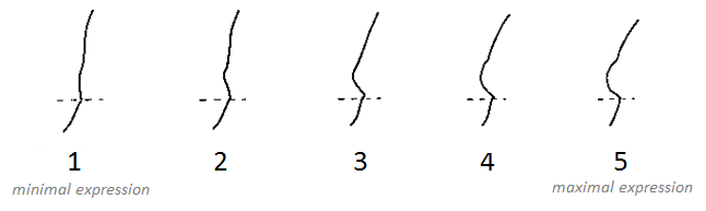
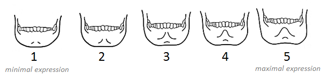
As with the five features of the skull used to sex a skeleton in the previous, the greater sciatic notch has also been given a numerical score from 1 to 5 relating to the level of expression. It has been generally found that female os coxae are more likely to exhibit a lower level of expression, whereas male os coxae are more likely to have higher levels of expression.

Figure 6:

Figure 6: It has been generally found that female os coxae are more likely to exhibit a lower level of expression, whereas male os coxae are more likely to have higher levels of expression, when it comes to the greater sciatic notch.

To obtain the best results whist examining the os coxae, it should be held in the same orientation as the pictured above. This allows you to match the angle of the greater sciatic to the closest expression that represents it. It should be noted that this method is usually used as a secondary indicator.

References:

Buikstra, J.E., Ubelaker, D.H. 1994. Standards for Data Collection From Human Skeletal Remains. Fayetteville, Arkansas: Arkansas Archaeological Survey Report Number 44.

Ubelaker, D.H. 1989. Human Skeletal Remains: Excavation, Analysis, Interpretation (2nd Ed.). Washington, DC: Taraxacum.

White, T.D., Folkens, P.A. 2005. The Human Bone Manual. San Diego, CA: Academic Press. Pg 392-398.

This is the third post of the Quick Tips series on sex determination of skeletal remains. The next post in this series will focus on the use of DNA to determine biological sex. To read more Quick Tips in the meantime, click here

Quick Tips: How To Estimate The Biological Sex Of A Human Skeleton – Skull Method.

This is the 2nd blog post in this Quick Tips series on estimating the biological sex of human skeletal remains. If you haven’t read the first post on the basics of sexing skeletal remains, click here to start at the beginning.

One of the most widely used methods of sexing skeletal remains is by examining the skull. The skull has five different features that are observed and scored.  The five features are the:

Markers together

Each of these markers is given a numerical score from 1 to 5 relating to the level of expression, with 1 being minimal expression and 5 being maximal expression. Each feature should be scored independently, and without influence from the other identifying features. It has been generally found that female skulls are more likely to have a lower level of expression in all features, whereas male skulls are more likely to have higher levels of expression.

To observe the nuchal crest, one should view the skull from its lateral profile and feel for the smoothness (1-minimal expression) or ruggedness (5-maximal expression) of the occipital surface, and compare it with the scoring system of that feature (Figure 1).

The scoring system for expression levels in the nuchal crest.

Figure 1: The scoring system for expression levels in the nuchal crest.

To observe the mastoid process, one should view the skull from its lateral profile and compare its size and volume, not its length, with other features of the skull such as the zygomatic process of the temporal lobe and external auditory meatus. Visually compare its size with the scoring system of that feature (Figure 2). If the mastoid process only descend or projects only a small distance then it should be scored a 1 (minimal expression), where as if it is several times the width and length of the external auditory meatus, then it should be scored as a 5 (maximal expression).

Figure 2: The scoring system for the size and volume of the mastoid process.

Figure 2: The scoring system for the expression levels of the mastoid process.

To observe the supraorbital margin, one should view the skull at it’s lateral profile and place their finger against the margin of the orbit and hold the edge to determine it’s thickness. If the edge feels ‘extremely sharp’ then it would score a 1minimal expression, if it felt rounded and thick as a pencil it would score a 5maximal expression (Figure 3).

Supraorbital Margin

Figure 3: The scoring system for the expression levels of the supraorbital margin.

To observe the supraorbital ridge, one should view the skull from it’s profile and view the prominence of the supraorbital ridge. If the ridge is smooth with little or no projection, then it would score a 1minimal expression, if it is pronounced and forms a rounded ‘loaf-shaped’ ridge then it would score a 5maximal expression (Figure 4).

Supraorbital Ridge - Glabella

Figure 4: The scoring system for the expression levels of the supraorbital ridge.

To observe the mental eminence, one should view the skull front facing, and hold the mandible between the thumbs and index fingers, with the thumbs placed either side of the mental eminence. If there is little or no projection of the mental eminence, then it would score a 1minimal expression, if it is pronounced it would score a 5maximal expression (Figure 5).

Mental Eminence

Figure 5: The scoring system for the expression levels of the mental eminence.

References:

Buikstra, J.E., Ubelaker, D.H. 1994. Standards for Data Collection From Human Skeletal Remains. Fayetteville, Arkansas: Arkansas Archaeological Survey Report Number 44.

Ubelaker, D.H. 1989. Human Skeletal Remains: Excavation, Analysis, Interpretation (2nd Ed.). Washington, DC: Taraxacum.

White, T.D., Folkens, P.A. 2005. The Human Bone Manual. San Diego, CA: Academic Press. Pg 360-385.

This is the second post of the Quick Tips series on sex determination of skeletal remains. The next post in this series will focus on the use of the pelvis and parturition scars to determine biological sex. To read more Quick Tips in the meantime, click here

Quick Tips: How To Estimate The Biological Sex Of A Human Skeleton – The Basics.

Within anthropological and archaeological sciences, ‘sex’ refers to the biological sex of an individual, based on the chromosomal difference of XX being female, and XY being male. Whereas ‘gender’ refers to the socio-cultural differences placed on the biological differences. In recent times, the words ‘gender’ and sex’ have been used incorrectly as interchangeable words within this discipline.

Therefore, it is important to remember that the word ‘gender’ refers an aspect of a person’s social identity, whereas ‘sex’ refers to the person’s biological identity.

Sexual dimorphism as seen in the human skeleton is determined by the hormones that are produced by the body. There are numerous markers on a human skeleton which can provide archaeologists and anthropologists with an estimate sex of the deceased. The areas of the skeletal remains that are studied are the:

 If the skeletal marker listed above is a link, it means that I have already covered it in an individual blog post and can be found by following the link.

The two most commonly used skeletal markers that are observed by osteologists are the skull and pelvic bone, as these show the most extreme differences.

It is generally noted that female skeleton elements are characterized by being smaller in size and lighter in construction, whereas males have larger, robust elements. Due to normal individual variation, there will always be smaller, dainty males and larger, robust females. Therefore, it is always important to observe a variety of skeletal markers to come to an accurate determination.

It should be noted that it is a lot harder to reliably deduce a juvenile/sub-adult’s sex, as many of the differences in skeletal markers only become visible after maturation, when the skeletal changes occur due to puberty. Therefore, use of DNA has been widely used to sex sub-adult skeletal remains as DNA analysis can now detect and identify X and Y chromosome-specific sequences.

References:

Buikstra, J.E., Ubelaker, D.H. 1994. Standards for Data Collection From Human Skeletal Remains. Fayetteville, Arkansas: Arkansas Archaeological Survey Report Number 44.

Ubelaker, D.H. 1989. Human Skeletal Remains: Excavation, Analysis, Interpretation (2nd Ed.). Washington, DC: Taraxacum.

White, T.D., Folkens, P.A. 2005. The Human Bone Manual. San Diego, CA: Academic Press. Pg 360-385.

This is the first of a Quick Tips series on sex determination of skeletal remains. The next post in this series will focus on the use of the skull to determine biological sex. To read more Quick Tips in the mean time, click here


Textbook of the Week: Archaeology in Practice.

Every week we highlight one archaeology/anthropology textbook from our suggested readings, a full list of our suggested resources can be found here, on our Useful Literature page.

ImageArchaeology in Practice: A Student Guide to Archaeological Analyses (UK/Europe Link)

Archaeology in Practice: A Student Guide to Archaeological Analyses (US/Worldwide Link)

by Jane Balme and Alistair Paterson. Rating: ****

“This is THE book for how to apply archaeological methods in real life contexts. It is easy to follow, so perfect for first year students as it uses numerous case studies and illustrations to show you how to apply it in practice. I used this during my studies to wrap my head around how methods can be applied – which helped when methods were only briefly discussed in theory during my lectures.”

If you’re new to the realm of archaeological, anthropological and forensic sciences (AAFS), or are a student needing sturdy and reliable references, or wondering “what archaeology or anthropology textbooks to buy? Check out our new ‘Useful Literature’ page!

Image

Textbook of the Week: Archaeology of Disease.

Every week we highlight one archaeology/anthropology textbook from our suggested readings, a full list of our suggested resources can be found here, on our Useful Literature page.

ImageThe Archaeology of Disease (UK/Europe)

The Archaeology of Disease (US/Worldwide Link)
(Click the links to have a peek inside the book)

by Charlotte Roberts and Keith Manchester. Rating: *****

“This is a essential if you’re studying diseases or taphonomy. It is fully illustrated with amazing case studies to display all diseases – ranging from simple fractures to malnutrition and infections.
Brilliant book, helped me a lot with my university Anthropology unit where I had to examine a bone and conclude which illness it had.”

If you’re new to the realm of archaeological, anthropological and forensic sciences (AAFS), or are a student needing sturdy and reliable references, or wondering “what archaeology or anthropology textbooks to buy? Check out our new ‘Useful Literature’ page!

Image

Textbook of the Week: Archaeology: Theories, Methods & Practice.

Every week we highlight one archaeology/anthropology textbook from our suggested readings, a full list of our suggested resources can be found here, on our Useful Literature page.

ImageArchaeology: Theories, Methods and Practice (UK/Europe Link)

Archaeology Essentials: Theories, Methods, and Practice (Second Edition) (US/Worldwide Link)

by Colin Renfrew and Paul Bahn. Rating: *****

“This book is highly acclaimed and is the ultimate archaeology bible for students, or people new to archaeology and want to get stuck right in. After being recommended it by two of my University lecturers, I took it out from the library to use for my assignments so many times I ended up buying it.”

If you’re new to the realm of archaeological, anthropological and forensic sciences (AAFS), or are a student needing sturdy and reliable references, or wondering “what archaeology or anthropology textbooks to buy? Check out our new ‘Useful Literature’ page!

Image