Quick Tips: How To Estimate The Biological Sex Of A Human Skeleton – The Basics.

Within anthropological and archaeological sciences, ‘sex’ refers to the biological sex of an individual, based on the chromosomal difference of XX being female, and XY being male. Whereas ‘gender’ refers to the socio-cultural differences placed on the biological differences. In recent times, the words ‘gender’ and sex’ have been used incorrectly as interchangeable words within this discipline.

Therefore, it is important to remember that the word ‘gender’ refers an aspect of a person’s social identity, whereas ‘sex’ refers to the person’s biological identity.

Sexual dimorphism as seen in the human skeleton is determined by the hormones that are produced by the body. There are numerous markers on a human skeleton which can provide archaeologists and anthropologists with an estimate sex of the deceased. The areas of the skeletal remains that are studied are the:

 If the skeletal marker listed above is a link, it means that I have already covered it in an individual blog post and can be found by following the link.

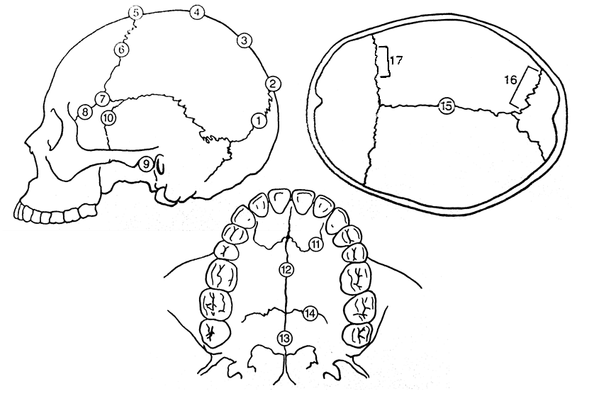
The two most commonly used skeletal markers that are observed by osteologists are the skull and pelvic bone, as these show the most extreme differences.

It is generally noted that female skeleton elements are characterized by being smaller in size and lighter in construction, whereas males have larger, robust elements. Due to normal individual variation, there will always be smaller, dainty males and larger, robust females. Therefore, it is always important to observe a variety of skeletal markers to come to an accurate determination.

It should be noted that it is a lot harder to reliably deduce a juvenile/sub-adult’s sex, as many of the differences in skeletal markers only become visible after maturation, when the skeletal changes occur due to puberty. Therefore, use of DNA has been widely used to sex sub-adult skeletal remains as DNA analysis can now detect and identify X and Y chromosome-specific sequences.

References:

Buikstra, J.E., Ubelaker, D.H. 1994. Standards for Data Collection From Human Skeletal Remains. Fayetteville, Arkansas: Arkansas Archaeological Survey Report Number 44.

Ubelaker, D.H. 1989. Human Skeletal Remains: Excavation, Analysis, Interpretation (2nd Ed.). Washington, DC: Taraxacum.

White, T.D., Folkens, P.A. 2005. The Human Bone Manual. San Diego, CA: Academic Press. Pg 360-385.

This is the first of a Quick Tips series on sex determination of skeletal remains. The next post in this series will focus on the use of the skull to determine biological sex. To read more Quick Tips in the mean time, click here


Quick Tips: How To Estimate The Chronological Age Of A Human Skeleton – Cranial Suture Closure Method.

This is the 4th blog post in this Quick Tips series on chronologically dating human skeletal remains, if you haven’t read the first post click here to start at the beginning. In my previous blog post I introduced the method of chronologically dating sub-adults using dentition, you can find out this information by clicking here.

Another method of chronologically aging human skeletal remains is by observing the cranial suture closure sites. The human skull has seventeen unique cranial fusion sites (Figure 1), that are positioned on the vault, the lateral-anterior sites, and the maxillary suture. The seventeen sites are:

  1. Midlambdoid                                           10.Superior sphenotemporal
  2. Lambda                                                    11. Incisive suture
  3. Obelion                                                    12. Anterior median palatine
  4. Anterior sagittal                                      13. Posterior median palatine
  5. Bregma                                                    14. Transverse palatine
  6. Midcoronal                                              15. Sagittal (endocranial)
  7. Pterion                                                     16. Left lambdoidal (endocranial)
  8. Sphenofrontal                                         17.Left coronal (endocranial)
  9. Inferior sphenotemporal
Image

Figure 1) Diagram showing the seventeen cranial suture sites.

The first seven fusion sites are on the vault, and the lateral-anterior sites consist of numbers six to ten. Each suture is usually given a numerical score, the score of 0-3 is recommended by the Buikstra and Ubelaker standards (1994). The Buikstra and Ubelaker (1994) scoring system is as follows;

  • 0 is given when the suture is open, meaning there is no evidence of ectocranial closure.
  • 1 is given where there is a minimal closure of the suture.
  • 2 is given to sutures with evidence of significant closure.
  • 3 is given to a completely obliterated suture (complete fusion).

So to attain the age of a skeletal remain you would total the scores for each grouping of sites, vault (1-7) or lateral anterior (6-10), and by comparing the scores to the known composite scores vs. chronological age of Meindl And Lovejoy, 1985 (Figure 2).

Image

Figure 2: Table demonstrating Meindl and Lovejoy (1985)’s composite scores of the sutures on the vault and lateral-anterior, respectively, in relation to mean chronological age.

A very useful cranial suture site is the sphenooccipital synchrondrosis, because at least 95% of all individuals have fusion here between the ages of twenty and twenty-five, with most individuals experiencing complete fusion around the age of twenty-three (Krogman and Işcan, 1986).

References:

Buikstra, J.E., Ubelaker, D.H. 1994. Standards for Data Collection From Human Skeletal Remains.Fayetteville, Arkansas: Arkansas Archaeological Survey Report Number 44.

Krogman, W.M., Işcan, M.Y. 1986. The Human Skeleton in Forensic Medicine (2nd Ed). Springfield, Illinois: C.C. Thomas.

Meindl, R.S., Lovejoy, C.O. 1985. Ectocranial Suture Closure: A Revised Method For The Determination Of Skeletal Age At Death Based On The Lateral-Anterior Sutures. American Journal of Physical Anthropology. 68, 57-66.

White, T.D., Folkens, P.A. 2005. The Human Bone Manual. San Diego, CA: Academic Press. Pg 360-385.

This is the forth of a Quick Tips series on ageing skeletal remains, the next in this series will focus on the use of the pubic symphyseal surface to chronologically age skeletal remains. To read more Quick Tips in the meantime, click here

To learn about basic fracture types and their characteristics/origins in their own Quick Tips series, click here!

Share Post Sign

Quick Tips: How To Estimate The Chronological Age Of A Human Skeleton – Epiphyseal Closure Method.

This Quick Tips post is the second in the series on age estimation on skeletal remains, if you haven’t read the previous post click here. The previous post provides an overview of the different techniques utilised by archaeologists/anthropologists, which will each be covered in more detail in their own blog post, and the categories that human skeletal remains are placed under according to their chronological age.

One of the methods frequently used by archaeologists/anthropologists to estimate the chronological age of human remains is by studying the level of epiphyseal fusions.

But first what is an epiphysis? An epiphysis is the cap at the end of a long bone that develops from a secondary ossification center. Over the course of adolescence the epiphysis, which is originally separate, will fuse to the diaphysis. The ages of which epiphyseal fusion begins and ends are very well documented, with the majority of epiphyseal activity taking place between the ages of fifteen and twenty-three.

Epiphyses

Diagram showing where the epiphysis is found.

As epiphyseal fusions are progressive they are often scored as either being unfused (non-union), united, and fully fused (complete union). Females often experience the union of many osteological elements before males, and every individual experience epiphyseal union at different ages.

Left: Diagram of a skeleton showing the position of the different epiphyseal elements. Right: A graph displaying the timing of fusion of epiphyses for various for various human osteological elements. The grey horizontal bars depict the period of time, in ages, when the fusion is occurring. All of the data is representative of males, except where it is noted. Data taken from Buikstra & Ubelaker, 1994.

Left: Diagram of a skeleton showing the position of the different epiphyseal elements.
Right: A graph displaying the timing of fusion of epiphyses for various for various human osteological elements. The grey horizontal bars depict the period of time, in ages, when the fusion is occurring. All of the data is representative of males, except where it is noted. Data taken from Buikstra & Ubelaker, 1994.

Archaeologists/anthropologists use standards that are well known and documented, such as Buikstra & Ubelaker’s (1994) depicted in the above graph. From the above data we know that, for example, the fusion of the femur head to the lesser trochanter is begins around the age of fifteen and a half and ends around the age of twenty. So if a skeleton has evidence of an unfused femur head/lesser trochanter, there is a possibility of the skeleton having a chronological age of < fifteen years. If there is full union of the epiphyses then the skeleton is more than likely being > twenty years old. But it should be noted that individuals vary in their development so numerous elements should be examined before coming to an accurate conclusion.

Different stages of epiphysis fusion of human tibias. Ages left to right: Newborn, 1.6 years old, six years old, ten years old, twelve years old and eighteen years old.

Different stages of epiphysis fusion of human tibias. Ages left to right: Newborn, 1.6 years old, six years old, ten years old, twelve years old and eighteen years old.

As several elements of the human skeleton begin the stages of epiphyseal fusion alongside the conclusion of tooth eruption, these two techniques (dentition and epiphyseal closure) are often used complementary to each other to help age sub-adults. The next post in this series on age estimation will focus on the use of dentition to aid with the chronological ageing of human remains.

References:

Buikstra, J.E., Ubelaker, D.H. 1994. Standards for Data Collection From Human Skeletal Remains. Fayetteville, Arkansas: Arkansas Archaeological Survey Report Number 44.

White, T.D., Folkens, P.A. 2005. The Human Bone Manual. San Diego, CA: Academic Press. Pg 360-385.

This is the second of a Quick Tips series on ageing skeletal remains, the next in this series will focus on the dentition method of ageing sub-adults. To read more Quick Tips in the mean time, click here

To learn about basic fracture types and their characteristics/origins click here!