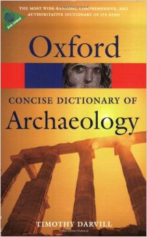
Textbook of the Week: Concise Oxford Dictionary of Archaeology.

Every week we highlight one archaeology/anthropology textbook from our suggested readings, a full list of our suggested resources can be found here, on our Useful Literature page.

51uiSHk+esL._SY344_BO1,204,203,200_

Concise Oxford Dictionary of Archaeology (UK/Europe Link)
Concise Oxford Dictionary of Archaeology  (US/Worldwide Link)
by Timothy Darvill. Rating: *****

“Does exactly as the title says – it is very concise, making it handy to have for your written assignments or dissertations, especially when you’re having trouble with interpretations of archaeological features! It is also useful to decipher what is being said within published papers by breaking down the archaeological jargon into layman terms if you’re a beginner!

If you’re a student – check out our ‘Quick Tips’ posts where we breakdown topics of AAFS into bite-sized chunks. We’re currently covering how to age and how to estimate the biological sex of skeletal remains, and also how to identify dental diseases!

Quick Tips: Identifying Dental Diseases – Dental Caries.  

Quick Tips: Identifying Dental Diseases – Dental Caries.  

In our previous Quick Tip post on identifying dental diseases, we gave a basic overview on the disease dental/enamel hypoplasia. If you haven’t read it, you can find it by clicking here.

Dental caries, also known as tooth decay, is thought to be the most common of dental diseases. This is due to it being recorded within archaeological populations more frequently than other dental diseases. It is an infectious and spreadable disease, which is the result of the fermentation of carbohydrates by bacteria that are present within teeth plaque. Its appearance can sometimes be observed as small opaque spots on the crowns of teeth, to large gaping cavities.

dental caries

Dental caries appearance can sometimes be observed as small opaque spots on the crowns of teeth, to large gaping cavities.

Dental caries occurs when sugars from the diet, particularly sucrose, are fermented by the bacteria Lactobacilus acidophilus and Streptococcys mutans, which are found within the built up plaque. This fermentation process causes acids to be produced, which in turn break down and demineralises teeth leaving behind cavities.

Powell (1985) divided the causes of dental caries into different areas, which are;

  • Environmental factors, the trace elements in food and water (i.e fluoride in water sources may protect against caries).
  • Pathogenic factors, the bacterial causing the disease.
  • Exogenous factors, from diet and oral hygiene.
  • Endogenous factors, the shape and structure of teeth.

Any part of the tooth structure that allows the accumulation of plaque and food debris can be susceptible to caries. This means that the crowns of the tooth (especially with molars and premolars due to the fissures), and the roots of the teeth are the areas most commonly affected by dental caries.

References:

Lukacs, J.R. 1989. Dental paleopathology: methods for reconstructing dietary patterns. In M.Y. Iscan and K.A.R. Kennedy (eds), Reconstruction of life from the skeleton. New York, Alan Liss, pp. 261-86.

Powell, M.L. 1985. The analysis of dental wear and caries for dietary reconstruction. In R.I. Gilbert and J.H. Mielke (eds), Analysis of prehistoric diets. London, Academic Press, pp. 307-38.

Ubelaker, D.H. 1989. Human Skeletal Remains: Excavation, Analysis, Interpretation (2nd Ed.). Washington, DC: Taraxacum.

White, T.D., Folkens, P.A. 2005. The Human Bone Manual. San Diego, CA: Academic Press. Pg 392-398.

This is the second post of the Quick Tips series on identifying dental diseases. The next post in this series will focus on how to identify calculus (calcified plague), and highlight the cause of this dental disease. To read more Quick Tips in the meantime, click here.

If you’re new to the realm of archaeological, anthropological and forensic sciences (AAFS), or are a student needing sturdy and reliable references, or wondering “what archaeology or anthropology textbooks are good? Check out our new ‘Useful Literature’ page for suggestions from peers and professors!
Quick Tips: Identifying Dental Diseases – Dental/Enamel Hypoplasia.

In our previous Quick Tip post on identifying dental diseases, we gave a basic overview on the different diseases that are observed. If you haven’t read it, you can find it by clicking here.

Dental hypoplasia is a condition that affects the enamel of a tooth. It is characterised by pits, grooves and transverse lines which are visible on the surface of tooth crowns. The lines, grooves and pits that are observed are defects in the enamels development. These defects occur when the enamel formation, also known as amelogenesis, is disturbed by a temporary stress to the organism which upsets the ameloblastic activity. Factors which can cause such stress and therefore disrupt the amelogenesis include; fever, malnutrition, and hypocalcemia.

Figure 1: An example of linear enamel hypoplasia.

Figure 1: An example of linear enamel hypoplasia.

It has been noted that enamel hypoplasia is more regularly seen on anterior teeth than on molars or premolars, and that the middle and cervical portions of enamel crowns tend to show more defects than the incisal third. This is due to the amelogenesis beginning at the occlusal apex of each tooth crown and proceeding rootward, towards where the crown then meets the root at the cervicoenamel line.

Figure 2: Anatomy of a tooth. Note the top third is known as either the occlusal third if in molars, or the incisal third when the tooth is an incisor or canine.

Figure 2: Anatomy of a tooth. Note the top third is known as either the occlusal third if in molars, or the incisal third when the tooth is an incisor or canine.

By studying these incidents of enamel hypoplasia within a population sample, we can be provided with valuable information regarding patterns of dietary stress and disease that may have occurred within the community.

References:

Lukacs, J.R. 1989. Dental paleopathology: methods for reconstructing dietary patterns. In M.Y. Iscan and K.A.R. Kennedy (eds), Reconstruction of life from the skeleton. New York, Alan Liss, pp. 261-86.

Ubelaker, D.H. 1989. Human Skeletal Remains: Excavation, Analysis, Interpretation (2nd Ed.). Washington, DC: Taraxacum.

White, T.D., Folkens, P.A. 2005. The Human Bone Manual. San Diego, CA: Academic Press. Pg 392-398.

This is the second post of the Quick Tips series on identifying dental diseases. The next post in this series will focus on how to identify dental caries and highlight the cause of this dental disease.

To read more Quick Tips in the meantime click here, or to learn about basic fracture types and their characteristics/origins click here!

Quick Tips: How To Estimate The Biological Sex Of A Human Skeleton – The Basics.

Within anthropological and archaeological sciences, ‘sex’ refers to the biological sex of an individual, based on the chromosomal difference of XX being female, and XY being male. Whereas ‘gender’ refers to the socio-cultural differences placed on the biological differences. In recent times, the words ‘gender’ and sex’ have been used incorrectly as interchangeable words within this discipline.

Therefore, it is important to remember that the word ‘gender’ refers an aspect of a person’s social identity, whereas ‘sex’ refers to the person’s biological identity.

Sexual dimorphism as seen in the human skeleton is determined by the hormones that are produced by the body. There are numerous markers on a human skeleton which can provide archaeologists and anthropologists with an estimate sex of the deceased. The areas of the skeletal remains that are studied are the:

 If the skeletal marker listed above is a link, it means that I have already covered it in an individual blog post and can be found by following the link.

The two most commonly used skeletal markers that are observed by osteologists are the skull and pelvic bone, as these show the most extreme differences.

It is generally noted that female skeleton elements are characterized by being smaller in size and lighter in construction, whereas males have larger, robust elements. Due to normal individual variation, there will always be smaller, dainty males and larger, robust females. Therefore, it is always important to observe a variety of skeletal markers to come to an accurate determination.

It should be noted that it is a lot harder to reliably deduce a juvenile/sub-adult’s sex, as many of the differences in skeletal markers only become visible after maturation, when the skeletal changes occur due to puberty. Therefore, use of DNA has been widely used to sex sub-adult skeletal remains as DNA analysis can now detect and identify X and Y chromosome-specific sequences.

References:

Buikstra, J.E., Ubelaker, D.H. 1994. Standards for Data Collection From Human Skeletal Remains. Fayetteville, Arkansas: Arkansas Archaeological Survey Report Number 44.

Ubelaker, D.H. 1989. Human Skeletal Remains: Excavation, Analysis, Interpretation (2nd Ed.). Washington, DC: Taraxacum.

White, T.D., Folkens, P.A. 2005. The Human Bone Manual. San Diego, CA: Academic Press. Pg 360-385.

This is the first of a Quick Tips series on sex determination of skeletal remains. The next post in this series will focus on the use of the skull to determine biological sex. To read more Quick Tips in the mean time, click here


On Location: Durotriges Project, 2014 – Roman Burials.

Header

Today’s On Location is featuring the Durotriges Project, an excavation in Winterborne Kingston,  Dorset, UK, which is held by Bournemouth University.

This archaeological site shows significant Iron Age and Roman settlement, particularly with the Durotriges tribe and Roman interaction. The Durotriges Project has been a yearly excavation, starting in 2009, which has uncovered many archaeological features. Over the past five years, the site has unearthed an Iron Age banjo enclosure, two Roman villas, an Iron Age burial site and numerous storage pits.

During this years excavation they were able to uncover a Bronze Age food preparation area surrounded by storage pits, and a flint mine – which was used to source the materials needed to construct the settlement’s buildings. Within the flint mine, students discovered the skeletal remains of an 18 month old juvenile.

Food Prep area DBD2014

The Bronze Age food preparation area, defined by post holes with a hearth in the centre, that was discovered during the Durotriges Project 2014. A storage pit can be seen to the right.

photo 3

The flint mine that was discovered in Trench 2. Within this flint mine, students unearthed the skeletal remains of an 18 month old juvenile.

The Durotriges Project recently hit the news this year with its discovery of five Roman burials. The burials, thought to date between 350AD to 380AD, were uncovered within a 15 by 15m  square enclosure around 100m away from the previously found Roman villa.

Roman Burials Within Enclosure

The five Roman era burials, dated between 350AD and 380AD, which were discovered within a square enclosure.

The skeletal remains found within the five graves have been identified as two males and three females. The males and two females have been aged between 40-50 years old, and the other female aged between 80-90 years old. The individuals were buried within coffins, with evidence of them being clothed as eyelets from their shoes were discovered.

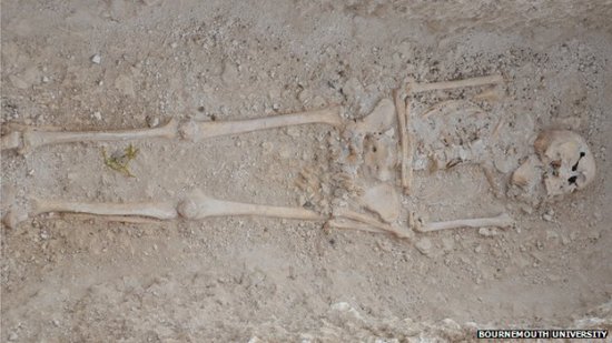
Skeleton

One of the five Roman burials uncovered by the Durotriges Project.

It is thought that they were of “high status” as were buried with great care and with burial gifts. It is hoped that the individuals were the occupants of the nearby Roman villa, and that their discovery will help shed light about the Roman’s diet, heath and ancestry.

To learn more about Bournemouth University’s Durotriges Project’s other fascinating discoveries from the site, you can view their blog here. Follow them on Twitter, and keep up to date with them by liking their Facebook page!