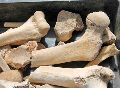
Quick Tips: How To Estimate The Chronological Age Of A Human Skeleton – Sternal Rib End Method.

This Quick Tips post is the sixth in the series on age estimation on skeletal remains, if you haven’t read the previous post click here, or to start at the beginning click here. The previous post provides an overview of the pubic symphyseal surface method of ageing, whereas the first post covers the basics.

The method was primarily developed by Iscan and Loth (1986) who studied the metamorphosis of the sternal end of the fourth rib. They found that the metamorphosis corresponds to the age but does vary by sex.

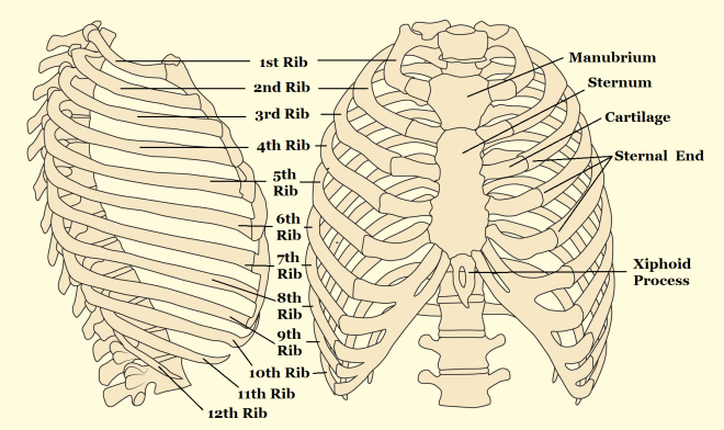
In their study they examined the “form, shape, texture and overall quality” of the sternal end which is found at the anterior (ventral) end of the shaft. This end is a roughened, porous, cupped oval surface which attaches to the cartilage attached to the sternum.  From this they were able to define a series of phases that depict the metamorphism of the sternal rib end over time.

Rib anatomy

Anatomy of the rib cage. This method was primarily developed by Iscan and Loth (1986) who studied the metamorphosis of the sternal end of the fourth rib. They found that the metamorphosis corresponds to the age but does vary by sex.

At the start the sternal end is flat or billowy with regular and rounded edges, and over time its rim thins and become irregular, with the surface porosity increasing, and the end becomes irregular. This method can be applied cautiously to the 3rd or 5th ribs as well, but not the others.

References:

Iscan, M.Y., and Loth, S.R. 1986. Estimation of age and determination of sex from the sternal rib. In: K. J. Reichs (ed.) Forensic Osteology: Advances in the Identification of Human Remains. Springfield, Illinois. Pg 68-89.

White, T.D., Folkens, P.A. 2005. The Human Bone Manual. San Diego, CA: Academic Press. Pg 360-385.

If you’re new to the realm of archaeological, anthropological and forensic sciences (AAFS), or are a student needing sturdy and reliable references, or wondering “what archaeology or anthropology textbooks to buy? Check out our new ‘Useful Literature’ page!


First Early Iron Age Burials Found In Dorset.

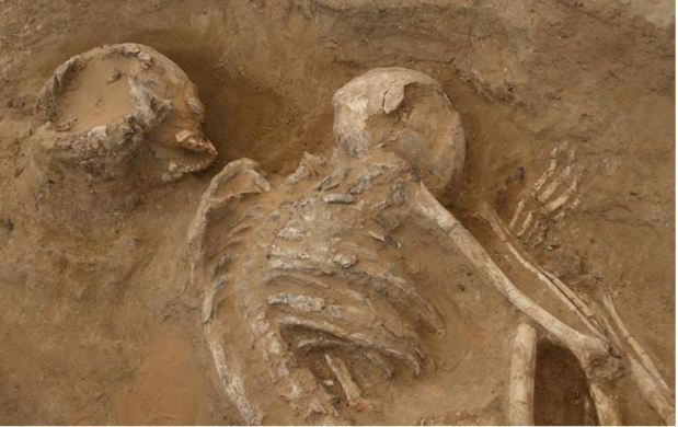
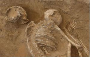
Archaeologists have discovered crouched burials of three skeletons, which are believed to be from the Bronze or Iron Age in Long Bredy, Dorset.  They were uncovered during a watching brief, being undertaken by the National Trust, on routine drainage works at an 18th century Dorset cottage.

The skeletal remains, thought to have been between eighteen and twenty five years old, have been radiocarbon dated which suggests they were buried between eight and six hundred BC. Due to the thick soils in the area, archaeologists tend to only stumble upon archaeological finds by accident whilst carrying out maintenance.

Dorset Crouched Burials

Archaeologists have discovered crouched burials of three skeletons, which are believed to be from the Bronze or Iron Age in Long Bredy, Dorset. © Martin Papworth

This find is the first burial from this time era that has been discovered in Dorset, making it a very significant find for the region. “There are no previous burials from that time in Dorset so it is a very significant find from the period with little evidence for the disposal of the dead,” says Martin Papworth, one of the archaeologists from the National Trust. “It’s an important window into the past, the first clues of the people who lived in Dorset at the time.”

To read the Unusual-ology post on the Ancient Egyptian use of lettuce as an aphrodisiac, click here, or how male spiders sacrifice themselves to their mate, click here. To learn about the recent vampire burials, and past vampire burials, click here.


Quick Tips: Archaeological Techniques –Use of Isotopes in Archaeology.

Isotopic analysis is widely used within the worlds of archaeology and anthropology. From analysing isotopes we’re able to uncover a wide range of information regarding the past; ranging from palaeoenvironments to palaeodiets, and even using isotopes to reconstruct trade routes of materials.

But first, what are isotopes?

All of the chemical elements consist of atoms which are specific to the element and the mass of an atom is dictated by the number of protons and neutrons it contains. The identity of the chemical element depends on the number of protons found within the atom’s nucleus, but the number of neutrons within the atom can vary. Atoms of the same chemical element (same number of protons), but with different masses, which is from the varying amount of neutrons, are called isotopes.

Stone Circle at Drombeg

Within nature, most of the elements consist of a number of isotopes. These isotopes can be found within water, livestock, crops and plants, which can then be used to reconstruct palaeodiets and palaeoenvironments.

Within nature, most of the elements consist of a number of isotopes. For a great majority of elements these relative proportions of isotopes are fixed, but there are a group of elements which either due to chemical or biochemical processes are of variable isotopic composition. These elements are oxygen, carbon, nitrogen and sulphur. Another group of isotopes that are used for analysis are strontium, lead and neodymium. These are formed by elements which contain stable but radiogenic isotopes, which are formed by radioactive decay of another element. Carbon and nitrogen isotope composition are primarily used to reconstruct diets, and oxygen isotopes are used to determine geographic origin. Strontium and lead isotopes found within teeth and bone can sometimes be used to reconstruct migration patterns in human populations and cultural affinity
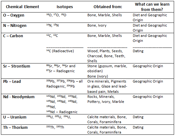
Isotopes Table

A table of the various elemental isotopes that are valuable in archaeological and anthropological research.

But how do isotopes get into skeletal remains?

Carbon isotopes are taken up through the diet of animals during their lifetime and these isotopes are deposited into teeth and bones of humans when they are consumed and digested. By studying animal bones and examining the 12C and 13C isotope ratio, it is possible to determine whether the animals ate predominately 3C or 4C plants. Oxygen isotopes are constantly being taken up and deposited into the body through the water a population drinks. This process ends with the organism’s death, from this point on isotopes no longer accumulate in the body, but do undergo degradation. For best result the researcher would need to know the original levels, or estimation thereof, of isotopes in the organism at the time of its death.

By creating a map of these natural occurring isotopes in different environments, rivers and areas, it is possible to identify where in an area the population lived, sourced their water or where the livestock grazed, by comparing the levels of isotopes that were obtained from skeletal remains to the environmental map. This mapping can also help identify trade routes that once existed and can also identify the migration patterns of populations.

References:

Balme, J., Paterson, A. 2006. Archaeology in Practice: A Student Guide to Archaeological Analayses. Oxford, UK: Blackwell Publishing. Pg 218.

Renfrew, C., Bahn, P. 1991. Archaeology: Theories, Methods and Practice. London, UK: Thames & Hudson. Pg 249-53.

If you’re new to the realm of archaeological, anthropological and forensic sciences (AAFS), or are a student needing sturdy and reliable references, or wondering “what archaeology or anthropology textbooks to buy? Check out our new ‘Useful Literature’ page!
Textbook of the Week: Flesh and Bone: An Introduction to Forensic Anthropology.

Every week we highlight one archaeology/anthropology textbook from our suggested readings, a full list of our suggested resources can be found here, on our Useful Literature page.

51dDAVU0ePL._SY344_BO1,204,203,200_

Flesh and Bone: An Introduction to Forensic Anthropology (UK/Europe)
Flesh and Bone: An Introduction to Forensic Anthropology (US/Worldwide Link)
by Miriam Nafte. Rating – *****
“This book was recommended to me by my professor – and was an absolute gem of a find! Perfect book for someone who wants an introduction to forensic anthropology, or has a single module in this topic.”

 If you’re a student – check out our ‘Quick Tips’ posts where we breakdown topics of AAFS into bite-sized chunks. We’re currently covering how to age and how to estimate the biological sex of skeletal remains, and also how to identify dental diseases!

Quick Tips – Common Questions: Why are some diseases more easily identified on skeletal remains than others?

This is a Quick Tips post providing a basic answer to a commonly asked question often faced within the field of archaeology and anthropology.

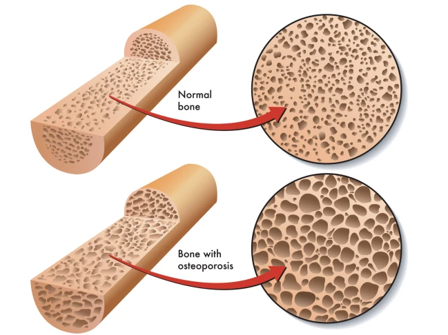
Some diseases are more easily to identify on skeletal remains due to leaving tell-tale signs in the bones preservation. An easy example of this is osteoporosis; this condition leaves the inners of bones a lot more porous which is easier to visually assess and compare to a ‘healthy’ individual’s skeletal remains.

canstockphoto18602582

Some diseases are more easily to identify on skeletal remains due to leaving tell-tale signs in the bones preservation. An  example of this is osteoporosis; this condition leaves the inners of bones a lot more porous than normal bones.

A study by Hershkovitz & Rothschild (1997) highlighted how certain medical conditions, in their study sickle cell anaemia, affects the bone growth and development. Hershkovitz & Rothschild found that due to the iron deficiency from sickle cell anaemia caused porotic hyperostosis (symmetrical osteoporosis) on the parietal bone as well as others. They were able to visually diagnose this due to the characteristic ‘pores’ over the skull.

Another example of an easily identifiable disease is tuberculosis (TB), TB can cause devastating bone damage. A recent archaeological study by Lewis (2011) looked into a population who suffered from TB. Lewis visually analysed the skeletal remains of a juvenile population from Poundbury Camp, Dorset. The TB infection caused numerous ailments to the infected, such as fever, but it’s the skeletal damage which gave the indication that the person suffered.  Amongst the population there was a high instance of skeletons with necrosis and lytic lesions characterised by minimal bone formation.  Many of the juvenile’s vertebrae displayed new bone formations which could indicate the presence of a paravertebral abscess. Many of the metatarsals were also displaying evidence of new bone formation which they concluded could be indicative of tuberculous dactylitis. Osteomyelitis, infection of the bone, was also found on a few mandibles and visually diagnosed due to its characteristic small pores found in a localised area. It is this characterised skeletal damage, seen on numerous cases during known TB outbreaks, which cause more diseases to be easily identified by eye due to the skeletal anomalies.

There are problems when trying to differentiate certain diseases for example; TB with brucellosis (undulant fever). As they both produce spinal lesions it is necessary to observe the other characteristic skeletal damage (new bone formation and osteomyelitis) to correctly identify it as a TB infection. Another slight difference between TB and brucellosis is that the spinal lesions are more sclerotic and regular than those from a TB infection (Lewis, 2011).

These porous bones and unexpected bone formations are easily observed, as they are not what’s expected during the known skeletal development found in healthy persons. Problems arise with diseases that do no damage to the skeleton, but instead affect soft tissue and muscles. These illnesses are harder to identify as they decay over time leaving only trace elements in the surrounding soils which would then hold the key for disease identification.

References:

Hershkovitz, I. Rothschild, B. et al. 1997. Recognition of sickle cell anemia in skeletal remains of children. American Journal of Physical Anthropology. Volume 104, Issue 2. 213-226.

Lewis, M. 2011. Tuberculosis in the non-adults from Romano-British Poundbury Camp, Dorset, England. International Journal of Paleopathology. Volume 1, Issue 1. 12-23.

To learn how archaeologists and anthropologists use teeth to age skeletal remains, read our Quick Tips: How To Estimate The Chronological Age of a Human Skeleton – Using Dentition to Age Subadults. Or to read more of our interesting Quick Tips, click here.

Unusual-ology: Strange 6,500-year-old Neolithic Burials Discovered in Egypt.

Unusual-ology: Strange 6,500-year-old Neolithic Burials Discovered in Egypt.

A team of archaeologists, led by Jacek Kabaciński from the Polish Academy of Sciences, have discovered the burials of sixty adults in a cemetery in Gebel Ramlah, Egypt.

Egypt Neolithic Burials

The unusual thing about these burials is the discovery of a grave that contained the skeletal remains of two individuals, one of which has deliberate cuts on their femur. These cut marks have not been seen in other Neolithic burials that have been unearthed in North Africa. But this particular grave wasn’t the only odd one they found. Kabaciński’s team found another two unusual graves one which was found to be lined with stone slabs, but it’s the third burial they discovered which is the oddest.

In the third grave, the skeletal remains of a man were found to be covered in pottery fragments, stones and lumps of red dye. Near his head a fragment of a Dorcas gazelle skull was found, which may have been used as a ceremonial headdress. The skeletal remains also showed signs of abnormal bone adhesions and fractures, leading Kabaciński to believe this man may have performed hunting rites.

Quick Tips: Identifying Dental Diseases – Dental Caries.  

Quick Tips: Identifying Dental Diseases – Dental Caries.  

In our previous Quick Tip post on identifying dental diseases, we gave a basic overview on the disease dental/enamel hypoplasia. If you haven’t read it, you can find it by clicking here.

Dental caries, also known as tooth decay, is thought to be the most common of dental diseases. This is due to it being recorded within archaeological populations more frequently than other dental diseases. It is an infectious and spreadable disease, which is the result of the fermentation of carbohydrates by bacteria that are present within teeth plaque. Its appearance can sometimes be observed as small opaque spots on the crowns of teeth, to large gaping cavities.

dental caries

Dental caries appearance can sometimes be observed as small opaque spots on the crowns of teeth, to large gaping cavities.

Dental caries occurs when sugars from the diet, particularly sucrose, are fermented by the bacteria Lactobacilus acidophilus and Streptococcys mutans, which are found within the built up plaque. This fermentation process causes acids to be produced, which in turn break down and demineralises teeth leaving behind cavities.

Powell (1985) divided the causes of dental caries into different areas, which are;

  • Environmental factors, the trace elements in food and water (i.e fluoride in water sources may protect against caries).
  • Pathogenic factors, the bacterial causing the disease.
  • Exogenous factors, from diet and oral hygiene.
  • Endogenous factors, the shape and structure of teeth.

Any part of the tooth structure that allows the accumulation of plaque and food debris can be susceptible to caries. This means that the crowns of the tooth (especially with molars and premolars due to the fissures), and the roots of the teeth are the areas most commonly affected by dental caries.

References:

Lukacs, J.R. 1989. Dental paleopathology: methods for reconstructing dietary patterns. In M.Y. Iscan and K.A.R. Kennedy (eds), Reconstruction of life from the skeleton. New York, Alan Liss, pp. 261-86.

Powell, M.L. 1985. The analysis of dental wear and caries for dietary reconstruction. In R.I. Gilbert and J.H. Mielke (eds), Analysis of prehistoric diets. London, Academic Press, pp. 307-38.

Ubelaker, D.H. 1989. Human Skeletal Remains: Excavation, Analysis, Interpretation (2nd Ed.). Washington, DC: Taraxacum.

White, T.D., Folkens, P.A. 2005. The Human Bone Manual. San Diego, CA: Academic Press. Pg 392-398.

This is the second post of the Quick Tips series on identifying dental diseases. The next post in this series will focus on how to identify calculus (calcified plague), and highlight the cause of this dental disease. To read more Quick Tips in the meantime, click here.

If you’re new to the realm of archaeological, anthropological and forensic sciences (AAFS), or are a student needing sturdy and reliable references, or wondering “what archaeology or anthropology textbooks are good? Check out our new ‘Useful Literature’ page for suggestions from peers and professors!

Quick Tips: Identifying Dental Diseases – Dental/Enamel Hypoplasia.

In our previous Quick Tip post on identifying dental diseases, we gave a basic overview on the different diseases that are observed. If you haven’t read it, you can find it by clicking here.

Dental hypoplasia is a condition that affects the enamel of a tooth. It is characterised by pits, grooves and transverse lines which are visible on the surface of tooth crowns. The lines, grooves and pits that are observed are defects in the enamels development. These defects occur when the enamel formation, also known as amelogenesis, is disturbed by a temporary stress to the organism which upsets the ameloblastic activity. Factors which can cause such stress and therefore disrupt the amelogenesis include; fever, malnutrition, and hypocalcemia.

Figure 1: An example of linear enamel hypoplasia.

Figure 1: An example of linear enamel hypoplasia.

It has been noted that enamel hypoplasia is more regularly seen on anterior teeth than on molars or premolars, and that the middle and cervical portions of enamel crowns tend to show more defects than the incisal third. This is due to the amelogenesis beginning at the occlusal apex of each tooth crown and proceeding rootward, towards where the crown then meets the root at the cervicoenamel line.

Figure 2: Anatomy of a tooth. Note the top third is known as either the occlusal third if in molars, or the incisal third when the tooth is an incisor or canine.

Figure 2: Anatomy of a tooth. Note the top third is known as either the occlusal third if in molars, or the incisal third when the tooth is an incisor or canine.

By studying these incidents of enamel hypoplasia within a population sample, we can be provided with valuable information regarding patterns of dietary stress and disease that may have occurred within the community.

References:

Lukacs, J.R. 1989. Dental paleopathology: methods for reconstructing dietary patterns. In M.Y. Iscan and K.A.R. Kennedy (eds), Reconstruction of life from the skeleton. New York, Alan Liss, pp. 261-86.

Ubelaker, D.H. 1989. Human Skeletal Remains: Excavation, Analysis, Interpretation (2nd Ed.). Washington, DC: Taraxacum.

White, T.D., Folkens, P.A. 2005. The Human Bone Manual. San Diego, CA: Academic Press. Pg 392-398.

This is the second post of the Quick Tips series on identifying dental diseases. The next post in this series will focus on how to identify dental caries and highlight the cause of this dental disease.

To read more Quick Tips in the meantime click here, or to learn about basic fracture types and their characteristics/origins click here!

3-Million Year Old Fossilised Metacarpals Show Evidence of Tool Use.

A recent study has put forward some important evidence of early human ancestors, in particular Australopithecus africanus, wielding tools in a human like fashion dating around 3 to 2-million years ago.

Figure 1: A recent study has put forward some important evidence of early human ancestors, in particular Australopithecus africanus (pictured), wielding tools in a human like fashion dating around 3 to 2-million years ago.

Figure 1: A recent study has put forward some important evidence of early human ancestors, in particular Australopithecus africanus (pictured), wielding tools in a human like fashion dating around 3 to 2-million years ago . ©Shaen Adey, Gallo Images/Corbis.

The study, led by Matthew Skinner from the University of Kent, compared the internal structures of the hand bones from the Australopithecus africanus and several Pleistocene hominins, which were previously considered to have not engaged in habitual tool use.

Skinner et al, found that they all have a human trabecular (spongy) bone pattern in the metacarpals, and this is consistent with the “forceful opposition of the thumb and fingers typically adopted during tool use”.

Top row: First metacarpals of the  various hominins.  Bottom row: 3-D renderings from the micro-CT scans showing a cross-section of the bone structure inside.

Figure 2: Top row: First metacarpals of the various hominids.
Bottom row: 3-D renderings from the micro-CT scans showing a cross-section of the bone structure inside. ©T.L. Kivell

The evolution of the hand, mainly the development of opposable thumbs, has been hailed as the key to success for early humans. It is thought that without the improvement of our grip and hand posture, tool technology could not have emerged and developed as well as it has.

This piece of research will provide a new discussion into when the first appearance of habitual tool use occurred in prehistory, as this study’s evidence of modern human-like tool use is dated 0.5-million years earlier than the first archaeological evidence of stone tools.

References:

Skinner, M. Stephens, N. Tsegai, Z. Foote, A. Nguyen, N. Gross, T. Pahr, D. Hublin, J. Kivell, T. 2015. Human-like hand use in Australopithecus africanusScience. 347, 6220. p395-399.
You can view this paper by clicking here.


If you’re a student – check out our ‘Quick Tips’ posts where we breakdown topics of AAFS into bite-sized chunks. We’re currently covering how to age and how to estimate the biological sex of skeletal remains, and also how to identify a variety of fracture types