Quick Tips: How To Estimate The Chronological Age Of A Human Skeleton – Sternal Rib End Method.

This Quick Tips post is the sixth in the series on age estimation on skeletal remains, if you haven’t read the previous post click here, or to start at the beginning click here. The previous post provides an overview of the pubic symphyseal surface method of ageing, whereas the first post covers the basics.

The method was primarily developed by Iscan and Loth (1986) who studied the metamorphosis of the sternal end of the fourth rib. They found that the metamorphosis corresponds to the age but does vary by sex.

In their study they examined the “form, shape, texture and overall quality” of the sternal end which is found at the anterior (ventral) end of the shaft. This end is a roughened, porous, cupped oval surface which attaches to the cartilage attached to the sternum.  From this they were able to define a series of phases that depict the metamorphism of the sternal rib end over time.

Rib anatomy

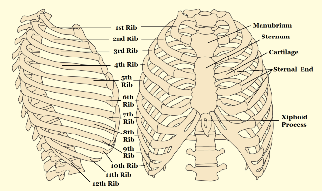
Anatomy of the rib cage. This method was primarily developed by Iscan and Loth (1986) who studied the metamorphosis of the sternal end of the fourth rib. They found that the metamorphosis corresponds to the age but does vary by sex.

At the start the sternal end is flat or billowy with regular and rounded edges, and over time its rim thins and become irregular, with the surface porosity increasing, and the end becomes irregular. This method can be applied cautiously to the 3rd or 5th ribs as well, but not the others.

References:

Iscan, M.Y., and Loth, S.R. 1986. Estimation of age and determination of sex from the sternal rib. In: K. J. Reichs (ed.) Forensic Osteology: Advances in the Identification of Human Remains. Springfield, Illinois. Pg 68-89.

White, T.D., Folkens, P.A. 2005. The Human Bone Manual. San Diego, CA: Academic Press. Pg 360-385.

If you’re new to the realm of archaeological, anthropological and forensic sciences (AAFS), or are a student needing sturdy and reliable references, or wondering “what archaeology or anthropology textbooks to buy? Check out our new ‘Useful Literature’ page!


Quick Tips: How To Estimate The Chronological Age Of A Human Skeleton – Cranial Suture Closure Method.

This is the 4th blog post in this Quick Tips series on chronologically dating human skeletal remains, if you haven’t read the first post click here to start at the beginning. In my previous blog post I introduced the method of chronologically dating sub-adults using dentition, you can find out this information by clicking here.

Another method of chronologically aging human skeletal remains is by observing the cranial suture closure sites. The human skull has seventeen unique cranial fusion sites (Figure 1), that are positioned on the vault, the lateral-anterior sites, and the maxillary suture. The seventeen sites are:

  1. Midlambdoid                                           10.Superior sphenotemporal
  2. Lambda                                                    11. Incisive suture
  3. Obelion                                                    12. Anterior median palatine
  4. Anterior sagittal                                      13. Posterior median palatine
  5. Bregma                                                    14. Transverse palatine
  6. Midcoronal                                              15. Sagittal (endocranial)
  7. Pterion                                                     16. Left lambdoidal (endocranial)
  8. Sphenofrontal                                         17.Left coronal (endocranial)
  9. Inferior sphenotemporal
Image

Figure 1) Diagram showing the seventeen cranial suture sites.

The first seven fusion sites are on the vault, and the lateral-anterior sites consist of numbers six to ten. Each suture is usually given a numerical score, the score of 0-3 is recommended by the Buikstra and Ubelaker standards (1994). The Buikstra and Ubelaker (1994) scoring system is as follows;

  • 0 is given when the suture is open, meaning there is no evidence of ectocranial closure.
  • 1 is given where there is a minimal closure of the suture.
  • 2 is given to sutures with evidence of significant closure.
  • 3 is given to a completely obliterated suture (complete fusion).

So to attain the age of a skeletal remain you would total the scores for each grouping of sites, vault (1-7) or lateral anterior (6-10), and by comparing the scores to the known composite scores vs. chronological age of Meindl And Lovejoy, 1985 (Figure 2).

Image

Figure 2: Table demonstrating Meindl and Lovejoy (1985)’s composite scores of the sutures on the vault and lateral-anterior, respectively, in relation to mean chronological age.

A very useful cranial suture site is the sphenooccipital synchrondrosis, because at least 95% of all individuals have fusion here between the ages of twenty and twenty-five, with most individuals experiencing complete fusion around the age of twenty-three (Krogman and Işcan, 1986).

References:

Buikstra, J.E., Ubelaker, D.H. 1994. Standards for Data Collection From Human Skeletal Remains.Fayetteville, Arkansas: Arkansas Archaeological Survey Report Number 44.

Krogman, W.M., Işcan, M.Y. 1986. The Human Skeleton in Forensic Medicine (2nd Ed). Springfield, Illinois: C.C. Thomas.

Meindl, R.S., Lovejoy, C.O. 1985. Ectocranial Suture Closure: A Revised Method For The Determination Of Skeletal Age At Death Based On The Lateral-Anterior Sutures. American Journal of Physical Anthropology. 68, 57-66.

White, T.D., Folkens, P.A. 2005. The Human Bone Manual. San Diego, CA: Academic Press. Pg 360-385.

This is the forth of a Quick Tips series on ageing skeletal remains, the next in this series will focus on the use of the pubic symphyseal surface to chronologically age skeletal remains. To read more Quick Tips in the meantime, click here

To learn about basic fracture types and their characteristics/origins in their own Quick Tips series, click here!

Share Post Sign