Quick Tips: Named Fractures – Part One: Hand & Forearms

This blog post is the 3rd in its series on bone fractures. To view the first blog post on the basic fracture types and information, including open and closed fractures, click here.

This blog post will highlight some of the common ‘named’ fractures you will often find in archaeological and anthropological settings. It is important to know their characteristics and common causes to help establish what happened – whether the fracture was received by defensive or offensive action, or purely accidental. This blog post will examine the first five common fractures associated with the hand and forearm bones.

Image

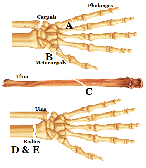
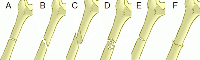
Common ‘Named’ Fractures of the forearms and hands: A) Boxer’s fracture, B) Bennett’s fractures, C) Parry’s or Monteggia’s fracture, D) Colles’ fractures, and E) Smith’s fractures.

The first two fractures we will look at affect the metacarpal bones;

A)     Boxer’s fracture: This fracture occurs due to the axial loading, meaning a force was applied along/parallel to the axis of the bone, on the transverse neck of the 4th and 5th metacarpal, secondary to an indirect force. A Boxer’s fracture often happens due to punching an object/person with a closed fist, hence the name ‘Boxer’ being associated to it.

B)      Bennett’s fracture: This fracture affects the 1st metacarpal (thumb) and extends into the carpometacarpal (CMC) joint which is complicated by subluxation (dislocation of a joint). A Bennett’s fracture is an oblique (See 1st blog post for meaning, click here) intra-articular metacarpal fracture caused by an axial force directed against the partially flexed metacarpal. This injury is also common when someone punches a hard object, but its most common cause is falling onto the thumb. An example of this is falling off a bike, as the thumb is extended around the handle bars.

The last three fractures affect the longbones of the forearm, the ulna and radius;

C)      Parry’s/Monteggia’s fracture: This fracture occurs on the proximal third of the ulna with subluxation of the radius/ulna. The most common cause of this fracture is by blunt force trauma caused by lifting the forearm up to protect the head or body in defence from an oncoming attack/striking object.

These two fractures affect the distal radius but cause displacement in two directions;

D)     Colles’ fracture: A Colles’ fracture, also known as a “dinner fork” or “bayonet” fracture, occurs when the distal radius is broken with dorsal displacement of the wrist and hand. This fracture is common when the person falls forwards and uses their outstretched hand to cushion the fall, which causes the force to displace and break the head of the radius.

E)      Smith’s fracture: A Smith’s fracture is the same as the Colles’ fracture but with ventral displacement of the broken radius head. The cause of a Smith’s fracture is the same as the Colles’ fracture, but it is less common.

Image

Fractures: D) Colles’ fracture, and E) Smith’s fracture.

The next Quick Tips post will discuss other ‘named fractures’ in archaeological/anthropological situations and their causes and characteristics.

This is the third post of a set on fractures, so keep your eyes open for the other posts, and the new ones to come. To view all the other Quick Tips posts click here!

Share Post Sign

Quick Tips: Fracture Types – The Basics Pt 2.

In my previous Quick Tips, which you can find by clicking here, you were introduced to the first six basic types of fractures, what the main causes of fractures are, and the two main categories they are classed in. It is important that you know the information in the previous Quick Tips post before learning these last few basic fractures, as it discussed the fundamentals of fractures.

Basic Fracture Types:

Image

Fracture Types: A) Butterfly, B) Longitudinal, C) Segmented, D) Hairline, and E) Avulsion.

A) Butterfly Fracture: Butterfly fractures usually affect long bones and can be caused by car accidents or by being knocked side on.

B) Longitudinal Fracture: As a transverse fracture is a bone along the horizontal axis, a longitudinal fracture is along the vertical axis.

C) Segmented Fracture: This is when the bone has fractures in two parts of the same bone causing the bone to break into larger bone fragments which are separated from the main body of a fractured bone.

D) Hairline Fracture: These fractures are also known as ‘stress fractures’. These types of fractures are very difficult to diagnose and once they heal there may be no evidence left to see. These are very difficult, if not impossible to identify when in an archaeological context.

E) Avulsion Fracture: Avulsion fractures are characterised as the separation of a small fragment of bone at the site of attachment of a ligament or tendon.

The next Quick Tips post will discuss the ‘named fractures’ that can be discovered in archaeological contexts, such as the familiar Bennett’s and Parry’s fracture, and their origins and common causes.

This post was put together by using knowledge from my degree and supplemented with the textbook ‘The Archaeology of Disease’ by Charlotte Roberts & Keith Manchester. If you’re interested in the latest scientific and archaeological techniques used to understand the diseases of past populations, you should check it out!

This is the second post of a set on fractures, so keep your eyes open for the other posts. To view other Quick Tips posts click here!

Quick Tips: Fracture Types – The Basics.

In my previous Quick Tips post I addressed how to distinguish between ante, peri and post-mortem fractures, click here if you haven’t read it yet. 

In this Quick Tips post I will show you some ways to identify and deduce common fracture types and their key characteristics. The definition of a fracture is a break in the continuity of a bone. There are three major causes of fractures: acute injury (an accident); underlying disease which then weakens the bone making it susceptible to fractures; and repeated stress (as seen in athletes).

All fracture types can be placed in two categories; open and closed. An open fracture, also known as a compound fracture, is where the bone breaks through the skin causing an open wound. It is called an open fracture as there is an open connection between the fracture site and skin. A closed fracture is where the bone has no connection between the outer skin surface and the fractured bone itself; it does not cause an open wound. A closed fracture is classed as a ‘simple fracture’.

Basic Fracture Types:

Image

Fracture Types: A) Transverse, B) Oblique, C) Spiral, D) Comminuted, E) Greenstick, and F) Impacted fracture.

A) Transverse Fracture: This is when the break of a fracture is in a horizontal line, it is the simplest fracture type.

B) Oblique Fracture: This fracture is a break which extends in a slanting direction.  Oblique fractures are caused by indirect or rotational force.

C) Spiral Fracture: As you can guess from the name, this is a fracture which is characterised by a spiral. It is often denoted as being caused by torsion or force onto the bone.

D) Comminuted Fracture: A comminuted fracture is characterised from the splintering of the bone. This causes the fracture to be made up of two or more pieces. This fracture is common in road-traffic accidents and these fractures are less likely to heal in a functionally satisfactory manner.

E) Greenstick Fracture: This occurs when a transverse fracture is incomplete. This fracture is seen mainly in children due to their young, immature bones which rarely break the whole width.

F) Impacted Fracture: This occurs when the bone is broken and not displaced but the two fractured ends are forced together. This produces a rather stable fracture which can heal readily but there may be some length lost.

This post was put together by using knowledge from my degree and supplemented with the textbook ‘The Archaeology of Disease’ by Charlotte Roberts & Keith Manchester. If you’re interested in the latest scientific and archaeological techniques used to understand the diseases of past populations, you should check it out!

This is the first post of a set, click here to read the second post. To view other Quick Tips posts click here!

Quick Tips: How can you tell if a skeletal fracture is ante, peri or post-mortem?

There is a relatively easy way to see whether a fracture to a skeleton is ante, peri or even post mortem. It is essential to detail and deduce which category a fracture falls into, as this is very important to see whether the fracture had played a part in the person’s death.

To first classify a fracture, we need to understand what the different categories mean. Some of you will already know these terminology, but here’s a quick reminder;

  • If a fracture is ante-mortem, it means that the fracture was made before death of the persons.
  • With peri-mortem fractures, it means that the fracture was received at or near the time of death of the persons – so could have been the fatal strike.
  • Post-mortem fractures are fractures that have been received after death, so during the time from death to the time of recovery. These fractures are usually from excavation processes, dismemberment, or even natural processes (soil, animal and plant activity).

You will be able to determine if a bone fracture was ante-mortem due to there being signs of healing which is shown by cell regrowth and repair.

With peri-mortem fractures, the person died before the healing started to take place, but the fractures will still contain the biomechanics that are present in ante-mortem fractures.

Post-mortem breaks tend to shatter compared to peri-mortem breaks which splinter, this is because bones which are in the post-mortem stage tend to be dry and rather brittle. Another big indicator of a fracture being post-mortem is the difference in colour.

The ‘Quick Tip’ that my applied anthropology lecturer taught me on how to easily distinguish between peri-mortem and post-mortem is to look at the fracture and decide; is it a clean break, as if you were breaking in half a bar of chocolate? If it is, then the fracture is most likely to be a peri-mortem fracture. If the break looks crumbly, like breaking a biscuit in half, it’s post-mortem fracture. Obviously this tip is not the most scientific, but it’s an easy way to begin your distinguishing process.

Image

Skull with signs of post-mortem fractures. This photo is from a practical lab session.

If you look at the photo above it illustrates a post-mortem fracture. You can determine this easily due to the colour difference on the edge of the fracture, where it is a much lighter colour compared to the rest of the skull and the crumbly nature of the cut.

References:

Most of this is my own knowledge that I learnt during my degree in my anthropology lectures/lab practical sessions. But if you’re looking for a published journal check the one below. It is very informative and easy to understand if you’re a beginner in the world of anthropology/archaeology! It also highlights some problems that can arise when distinguishing trauma, it’s really interesting!

Smith, A.C. 2010. Distinguishing Between Antemortem, Perimortem, and Postmortem Trauma. Academia.edu. Available from here in .pdf form!

Read more anthropology/archaeology quick tips here!