Quick Tips – Common Questions: Why are some diseases more easily identified on skeletal remains than others?

This is a Quick Tips post providing a basic answer to a commonly asked question often faced within the field of archaeology and anthropology.

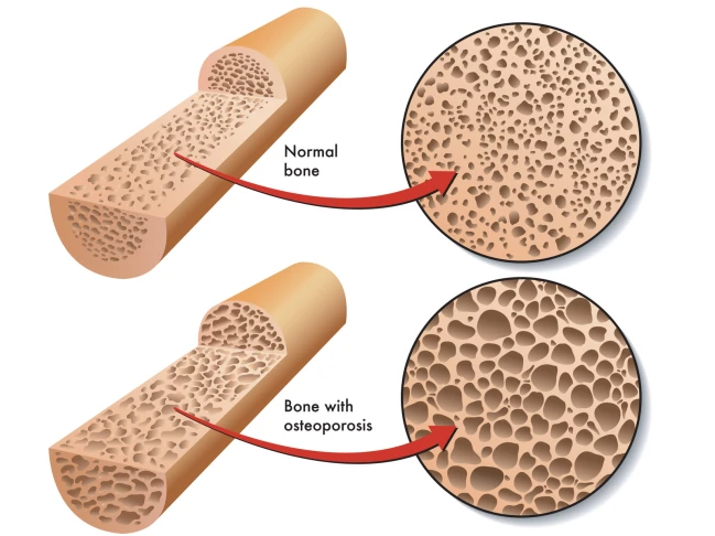
Some diseases are more easily to identify on skeletal remains due to leaving tell-tale signs in the bones preservation. An easy example of this is osteoporosis; this condition leaves the inners of bones a lot more porous which is easier to visually assess and compare to a ‘healthy’ individual’s skeletal remains.
canstockphoto18602582

Some diseases are more easily to identify on skeletal remains due to leaving tell-tale signs in the bones preservation. An  example of this is osteoporosis; this condition leaves the inners of bones a lot more porous than normal bones.

A study by Hershkovitz & Rothschild (1997) highlighted how certain medical conditions, in their study sickle cell anaemia, affects the bone growth and development. Hershkovitz & Rothschild found that due to the iron deficiency from sickle cell anaemia caused porotic hyperostosis (symmetrical osteoporosis) on the parietal bone as well as others. They were able to visually diagnose this due to the characteristic ‘pores’ over the skull.

Another example of an easily identifiable disease is tuberculosis (TB), TB can cause devastating bone damage. A recent archaeological study by Lewis (2011) looked into a population who suffered from TB. Lewis visually analysed the skeletal remains of a juvenile population from Poundbury Camp, Dorset. The TB infection caused numerous ailments to the infected, such as fever, but it’s the skeletal damage which gave the indication that the person suffered.  Amongst the population there was a high instance of skeletons with necrosis and lytic lesions characterised by minimal bone formation.  Many of the juvenile’s vertebrae displayed new bone formations which could indicate the presence of a paravertebral abscess. Many of the metatarsals were also displaying evidence of new bone formation which they concluded could be indicative of tuberculous dactylitis. Osteomyelitis, infection of the bone, was also found on a few mandibles and visually diagnosed due to its characteristic small pores found in a localised area. It is this characterised skeletal damage, seen on numerous cases during known TB outbreaks, which cause more diseases to be easily identified by eye due to the skeletal anomalies.

There are problems when trying to differentiate certain diseases for example; TB with brucellosis (undulant fever). As they both produce spinal lesions it is necessary to observe the other characteristic skeletal damage (new bone formation and osteomyelitis) to correctly identify it as a TB infection. Another slight difference between TB and brucellosis is that the spinal lesions are more sclerotic and regular than those from a TB infection (Lewis, 2011).

These porous bones and unexpected bone formations are easily observed, as they are not what’s expected during the known skeletal development found in healthy persons. Problems arise with diseases that do no damage to the skeleton, but instead affect soft tissue and muscles. These illnesses are harder to identify as they decay over time leaving only trace elements in the surrounding soils which would then hold the key for disease identification.

References:

Hershkovitz, I. Rothschild, B. et al. 1997. Recognition of sickle cell anemia in skeletal remains of children. American Journal of Physical Anthropology. Volume 104, Issue 2. 213-226.

Lewis, M. 2011. Tuberculosis in the non-adults from Romano-British Poundbury Camp, Dorset, England. International Journal of Paleopathology. Volume 1, Issue 1. 12-23.

To learn how archaeologists and anthropologists use teeth to age skeletal remains, read our Quick Tips: How To Estimate The Chronological Age of a Human Skeleton – Using Dentition to Age Subadults. Or to read more of our interesting Quick Tips, click here.

Quick Tips – Common Questions: Can physical activities undertaken during life be detected on skeletal remains?

This is a Quick Tips post providing a basic answer to a commonly asked question often faced within the field of archaeology and anthropology.

Can physical activities undertaken during life be detected on skeletal remains? Yes they can.

Numerous activities, such as hunting, gathering, exercise and more obviously fighting, can inflict damage or adaptations onto to a skeletal system. Some physical activities can be easily identified by due to the damage they can produce to the skeleton, i.e. fighting, whereas the skeletons adapt to strain caused by sport or a daily activity can be harder to detect.

Stock (2006) investigated hunter-gatherer postcranial robusticity relative to patterns of mobility and climatic adaption. In this study, Stock took four collections of known hunter-gatherers skeletal remains along with the associated data of the environmental factors in the population area and the terrestrial mobility. In every analysis conducted, the effective environmental temperature was found to be negatively correlated with strength. Stock concluded that hunter-gatherers from colder climates tend to have stronger long bone diaphysis, than the groups from warmer regions. Although in contrast, the partial correlations between mobility and robusticity are positive; suggesting that activity has a consistently positive relationship with diaphyseal strength. This study indicates that even the simple ‘easy’ activity of hunting and gathering can affect diaphyseal strength of a skeleton and that the activity can be detected.

Exercise is also one of the most common factors to cause a skeleton to adapt. A recent study by Shaw (2009) was able to correctly predict an athlete’s chosen sport from quantifying the soft tissue properties and bone morphology. In Shaw’s study he focused on examining modern athletes (runners, field hockey players, swimmers, and cricketers) and a control group. Using peripheral quantitative computed tomography (pQCT), Shaw quantified the relationship between the amount of muscle and other soft tissues and the morphology of the bones along the midshaft of the arm, forearm and lower legs. This study concluded that Shaw could correctly identify an athlete’s chosen sport from examining a skeletal system and quantifying the bone mass and strength. Shaw concluded that the changes to the bones structural properties were from the strain of daily habitual training from the athlete’s young age.

These two modern studies, Stock (2006) and Shaw (2009), perfectly highlight how physical activities can be detected on skeletal remains.  But these morphological changes can be harder to detect than more brutal activities such as fighting. This is because war and fights leave tell-tale marks on the skeletons which are detectable from eye rather than quantitating data. Violence within a population whether its ritual/habitual, in times of war or domestic can be easily identified from the fractures and dents a bone receives.

A recent NAI (Non-accidental Injury) study from Day et al (2006), highlighted how skeletal remains could indicate bone trauma caused by violence. The study retrospectively observed cases of suspected NAI injuries sustained by children from X-rays obtained at an Edinburgh hospital. The bone fractures, mostly found on the skull and long bones, were suspected to be cause by domestic abuse and evidence of blunt force trauma was observed in numerous cases. Even though this is a recent study conducted on NAI instances, it does appropriately show how violence can inflict damage onto skeletal remains. An archaeological skeleton could show healed/unhealed fractures sustained via a physically demanding activity which was violent in nature, such as war or ritual fighting.

References:

Day, F. Clegg, S. McPhillips, M. Mok, J. 2006. A retrospective case series of skeletal surveys in children with suspected non-accidental injury. Journal of Clinical Forensic Medicine. 13, 12. 55-59.

Shaw, C. 2009. ‘Putting flesh back onto the bones?’ Can we predict soft tissue properties from skeletal and fossil remains?. Journal of Human Evolution. 59, 5. 484-492.

Stock, J.T. 2006. Hunter-Gatherer Postcranial Robusticity Relative to Patterns of Mobility, Climatic Adaption and Selective Tissue Economy. American Journal of Physical Anthropology. 131, 2. 194-203.