Quick Tips: How To Estimate The Biological Sex Of A Human Skeleton – Skull Method.

This is the 2nd blog post in this Quick Tips series on estimating the biological sex of human skeletal remains. If you haven’t read the first post on the basics of sexing skeletal remains, click here to start at the beginning.

One of the most widely used methods of sexing skeletal remains is by examining the skull. The skull has five different features that are observed and scored.  The five features are the:

Markers together

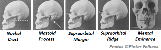
Each of these markers is given a numerical score from 1 to 5 relating to the level of expression, with 1 being minimal expression and 5 being maximal expression. Each feature should be scored independently, and without influence from the other identifying features. It has been generally found that female skulls are more likely to have a lower level of expression in all features, whereas male skulls are more likely to have higher levels of expression.

To observe the nuchal crest, one should view the skull from its lateral profile and feel for the smoothness (1-minimal expression) or ruggedness (5-maximal expression) of the occipital surface, and compare it with the scoring system of that feature (Figure 1).

The scoring system for expression levels in the nuchal crest.

Figure 1: The scoring system for expression levels in the nuchal crest.

To observe the mastoid process, one should view the skull from its lateral profile and compare its size and volume, not its length, with other features of the skull such as the zygomatic process of the temporal lobe and external auditory meatus. Visually compare its size with the scoring system of that feature (Figure 2). If the mastoid process only descend or projects only a small distance then it should be scored a 1 (minimal expression), where as if it is several times the width and length of the external auditory meatus, then it should be scored as a 5 (maximal expression).

Figure 2: The scoring system for the size and volume of the mastoid process.

Figure 2: The scoring system for the expression levels of the mastoid process.

To observe the supraorbital margin, one should view the skull at it’s lateral profile and place their finger against the margin of the orbit and hold the edge to determine it’s thickness. If the edge feels ‘extremely sharp’ then it would score a 1minimal expression, if it felt rounded and thick as a pencil it would score a 5maximal expression (Figure 3).

Supraorbital Margin

Figure 3: The scoring system for the expression levels of the supraorbital margin.

To observe the supraorbital ridge, one should view the skull from it’s profile and view the prominence of the supraorbital ridge. If the ridge is smooth with little or no projection, then it would score a 1minimal expression, if it is pronounced and forms a rounded ‘loaf-shaped’ ridge then it would score a 5maximal expression (Figure 4).

Supraorbital Ridge - Glabella

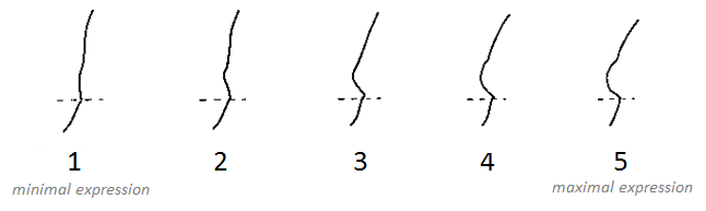
Figure 4: The scoring system for the expression levels of the supraorbital ridge.

To observe the mental eminence, one should view the skull front facing, and hold the mandible between the thumbs and index fingers, with the thumbs placed either side of the mental eminence. If there is little or no projection of the mental eminence, then it would score a 1minimal expression, if it is pronounced it would score a 5maximal expression (Figure 5).

Mental Eminence

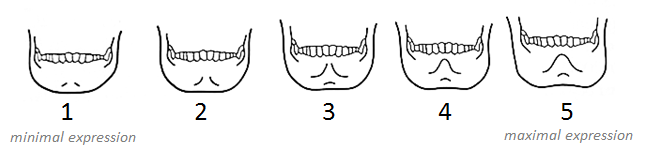
Figure 5: The scoring system for the expression levels of the mental eminence.

References:

Buikstra, J.E., Ubelaker, D.H. 1994. Standards for Data Collection From Human Skeletal Remains. Fayetteville, Arkansas: Arkansas Archaeological Survey Report Number 44.

Ubelaker, D.H. 1989. Human Skeletal Remains: Excavation, Analysis, Interpretation (2nd Ed.). Washington, DC: Taraxacum.

White, T.D., Folkens, P.A. 2005. The Human Bone Manual. San Diego, CA: Academic Press. Pg 360-385.

This is the second post of the Quick Tips series on sex determination of skeletal remains. The next post in this series will focus on the use of the pelvis and parturition scars to determine biological sex. To read more Quick Tips in the meantime, click here

Quick Tips: How To Estimate The Chronological Age Of A Human Skeleton – Using Dentition To Age Subadults.

This Quick Tips post is the third in the series on age estimation of human skeletal remains, if you haven’t read the first post click here to start at the beginning. The first post provides an overview of the different techniques utilised by archaeologists/anthropologists, which will each be covered in more detail in their own blog post, and the categories that human skeletal remains are placed under according to their chronological age. The second post examines the epiphyseal closure method, which you can find here.

The practice of using dentition to chronologically age human skeletal remains is split into two halves, depending on the whether the skeleton is that of a subadult or adult. This blog post is going to discuss using dentition to age subadults.

Due to the abundance of teeth found in many archaeological, forensic, paleontological, and anthropological contexts and because of the regular tooth formation and eruption times, dental development is the most widely used technique for aging subadult remains. As stated in my previous blog post, several elements of the human skeleton begin the stages of epiphyseal fusion alongside the conclusion of tooth eruption; these two techniques (dentition and epiphyseal closure) are often used complementary to each other to help age sub-adults. When it comes to subadult tooth emergence there are four stages:

Stage 1 is where most of the deciduous teeth, commonly referred to as ‘milk teeth’, emerge during the second year of life.

Stage 2, during this stage the two permanent incisors and the first permanent molar emerge, this stage typically occurs between the age of six and eight years.

Stage 3, occurs between the age of ten and twelve and it involves the emergence of the permanent canines, premolars, and second molars.

Stage 4, or the final stage involves the third molar emerging around the age of eighteen years.

When looking at dentition you must look at all aspects of emergence and not just at the fully erupted tooth, which includes the completeness of all roots and crowns (formation) and the position of each tooth relative to the alveolar margin (eruption). Ubelaker (1989) conducted a study on non-Native Americans and created a graphic summary of dental development and the correlating ages it occurs, see figure 1.

Figure 1: Ubelaker's (1989) diagram showing the dental development in correlation to age.

Figure 1: Ubelaker’s (1989) diagram showing the dental development in correlation to age.

It must be noted that the ages these stages occur at differ per individual so only act as a reference. Gustafson and Koch (1974) created a graph to illustrate the variation that could occur with dental development, see figure 2.

Figure 2: Gustafson and Koch's (1974) image showing the variation in timing of dental development. Colour key: Black highlights the age that crown mineralization begins, Dark grey shows the age of crown completion, Light grey shows the age of eruption, and White displays age of root completion.

Figure 2: Gustafson and Koch’s (1974) image showing the variation in timing of dental development. Colour key: Black highlights the age that crown mineralization begins, Dark grey shows the age of crown completion, Light grey shows the age of eruption, and White displays age of root completion.

References:

Gustafson, G. Koch, G. 1974. Age estimation up to 16 years of age based on dental development. Odontologisk Revy. 25. Pg 297-306.

Ubelaker, D.H. 1989. Human Skeletal Remains: Excavation, Analysis, Interpretation (2nd Ed.). Washington, DC: Taraxacum.

White, T.D., Folkens, P.A. 2005. The Human Bone Manual. San Diego, CA: Academic Press. Pg 360-385.

This is the third of a Quick Tips series on ageing skeletal remains, the next in this series will focus on the use of dentition to age adults and the use of cranial suture closure. To read more Quick Tips in the meantime, click here

To learn about basic fracture types and their characteristics/origins in their own Quick Tips series, click here!


Quick Tips: How To Estimate The Chronological Age Of A Human Skeleton – Epiphyseal Closure Method.

This Quick Tips post is the second in the series on age estimation on skeletal remains, if you haven’t read the previous post click here. The previous post provides an overview of the different techniques utilised by archaeologists/anthropologists, which will each be covered in more detail in their own blog post, and the categories that human skeletal remains are placed under according to their chronological age.

One of the methods frequently used by archaeologists/anthropologists to estimate the chronological age of human remains is by studying the level of epiphyseal fusions.

But first what is an epiphysis? An epiphysis is the cap at the end of a long bone that develops from a secondary ossification center. Over the course of adolescence the epiphysis, which is originally separate, will fuse to the diaphysis. The ages of which epiphyseal fusion begins and ends are very well documented, with the majority of epiphyseal activity taking place between the ages of fifteen and twenty-three.

Epiphyses

Diagram showing where the epiphysis is found.

As epiphyseal fusions are progressive they are often scored as either being unfused (non-union), united, and fully fused (complete union). Females often experience the union of many osteological elements before males, and every individual experience epiphyseal union at different ages.

Left: Diagram of a skeleton showing the position of the different epiphyseal elements. Right: A graph displaying the timing of fusion of epiphyses for various for various human osteological elements. The grey horizontal bars depict the period of time, in ages, when the fusion is occurring. All of the data is representative of males, except where it is noted. Data taken from Buikstra & Ubelaker, 1994.

Left: Diagram of a skeleton showing the position of the different epiphyseal elements.
Right: A graph displaying the timing of fusion of epiphyses for various for various human osteological elements. The grey horizontal bars depict the period of time, in ages, when the fusion is occurring. All of the data is representative of males, except where it is noted. Data taken from Buikstra & Ubelaker, 1994.

Archaeologists/anthropologists use standards that are well known and documented, such as Buikstra & Ubelaker’s (1994) depicted in the above graph. From the above data we know that, for example, the fusion of the femur head to the lesser trochanter is begins around the age of fifteen and a half and ends around the age of twenty. So if a skeleton has evidence of an unfused femur head/lesser trochanter, there is a possibility of the skeleton having a chronological age of < fifteen years. If there is full union of the epiphyses then the skeleton is more than likely being > twenty years old. But it should be noted that individuals vary in their development so numerous elements should be examined before coming to an accurate conclusion.

Different stages of epiphysis fusion of human tibias. Ages left to right: Newborn, 1.6 years old, six years old, ten years old, twelve years old and eighteen years old.

Different stages of epiphysis fusion of human tibias. Ages left to right: Newborn, 1.6 years old, six years old, ten years old, twelve years old and eighteen years old.

As several elements of the human skeleton begin the stages of epiphyseal fusion alongside the conclusion of tooth eruption, these two techniques (dentition and epiphyseal closure) are often used complementary to each other to help age sub-adults. The next post in this series on age estimation will focus on the use of dentition to aid with the chronological ageing of human remains.

References:

Buikstra, J.E., Ubelaker, D.H. 1994. Standards for Data Collection From Human Skeletal Remains. Fayetteville, Arkansas: Arkansas Archaeological Survey Report Number 44.

White, T.D., Folkens, P.A. 2005. The Human Bone Manual. San Diego, CA: Academic Press. Pg 360-385.

This is the second of a Quick Tips series on ageing skeletal remains, the next in this series will focus on the dentition method of ageing sub-adults. To read more Quick Tips in the mean time, click here

To learn about basic fracture types and their characteristics/origins click here!

Quick Tips: How To Estimate The Chronological Age Of A Human Skeleton – The Basics.

Estimation of age-at-death involves observing morphological features in the skeletal remains, comparing the information with changes recorded for recent populations of known age, and then estimating any sources of variability likely to exist between the prehistoric and the recent population furnishing the documented data. This third step is seldom recognized or discussed in osteological studies, but it represents a significant element. – Ubelaker, D. 1989.

There are numerous markers on a human skeleton which can provide archaeologists and anthropologists with an estimate age of the deceased. The areas of the skeletal remains that are studied are:

If the skeletal marker listed above is a link, it means that I have already covered it in an individual blog post and can be found by following the link.

We can age skeletal remains to a rough estimate, as over a lifetime a human skeleton undergoes sequential chronological changes. Teeth appear and bone epiphyseal form and fuse during childhood and adolescence, with some bone fusing, metamorphose and degeneration carrying on after the age of twenty. Buikstra and Ubelaker, 1994, developed seven age categories that human osteological remains are separated into. The seven age classes are; fetus (before birth), infant (0-3 years), child (3-12 years), adolescent (12-20 years), young adult (20-35 years), middle adult (35-50 years), and old adult (50+ years).

When it comes to ageing skeletal remains, there are numerous problems. This is because individuals of the same chronological age can show difference degrees of development. Therefore, this causes archaeologists and anthropologists to obtain an accurate age estimate, which may not be precise.

It should be noted that it is a lot easier to deduce a juvenile/sub-adult’s age, as the ends of the limb bones form and fuse at known ages and the ages of which tooth formation and eruption occur are very well documented, although somewhat variable. After maturity there is little continuing skeletal change to observe, this causes adult ageing to become more difficult.

References:

Buikstra, J.E., Ubelaker, D.H. 1994. Standards for Data Collection From Human Skeletal Remains. Fayetteville, Arkansas: Arkansas Archaeological Survey Report Number 44.

Ubelaker, D.H. 1989. Human Skeletal Remains: Excavation, Analysis, Interpretation (2nd Ed.). Washington, DC: Taraxacum.

White, T.D., Folkens, P.A. 2005. The Human Bone Manual. San Diego, CA: Academic Press. Pg 360-385.

This is the first of a Quick Tips series on ageing skeletal remains, the next in this series will focus on the epiphyseal closure method of ageing sub-adults. To read more Quick Tips in the mean time, click here

To learn about basic fracture types and their characteristics/origins click here!